

Neuronal ensembles: Building blocks of neural circuits
- PMID: 38262413
- PMCID: PMC10957317
- DOI: 10.1016/j.neuron.2023.12.008
Neuronal ensembles: Building blocks of neural circuits
Abstract
Neuronal ensembles, defined as groups of neurons displaying recurring patterns of coordinated activity, represent an intermediate functional level between individual neurons and brain areas. Novel methods to measure and optically manipulate the activity of neuronal populations have provided evidence of ensembles in the neocortex and hippocampus. Ensembles can be activated intrinsically or in response to sensory stimuli and play a causal role in perception and behavior. Here we review ensemble phenomenology, developmental origin, biophysical and synaptic mechanisms, and potential functional roles across different brain areas and species, including humans. As modular units of neural circuits, ensembles could provide a mechanistic underpinning of fundamental brain processes, including neural coding, motor planning, decision-making, learning, and adaptability.
Keywords: assembly; attractor; neural networks; synfire.
Copyright © 2023 Elsevier Inc. All rights reserved.
Conflict of interest statement
Declaration of interests The authors declare no competing interests.
Figures
Similar articles
-
Intrinsic excitability mechanisms of neuronal ensemble formation.Elife. 2022 May 4;11:e77470. doi: 10.7554/eLife.77470. Elife. 2022. PMID: 35506662 Free PMC article.
-
Identification of Pattern Completion Neurons in Neuronal Ensembles Using Probabilistic Graphical Models.J Neurosci. 2021 Oct 13;41(41):8577-8588. doi: 10.1523/JNEUROSCI.0051-21.2021. Epub 2021 Aug 19. J Neurosci. 2021. PMID: 34413204 Free PMC article.
-
Playing the piano with the cortex: role of neuronal ensembles and pattern completion in perception and behavior.Curr Opin Neurobiol. 2020 Oct;64:89-95. doi: 10.1016/j.conb.2020.03.014. Epub 2020 Apr 10. Curr Opin Neurobiol. 2020. PMID: 32320944 Free PMC article. Review.
-
Imaging and Optically Manipulating Neuronal Ensembles.Annu Rev Biophys. 2017 May 22;46:271-293. doi: 10.1146/annurev-biophys-070816-033647. Epub 2017 Mar 15. Annu Rev Biophys. 2017. PMID: 28301770 Review.
-
Creation of Neuronal Ensembles and Cell-Specific Homeostatic Plasticity through Chronic Sparse Optogenetic Stimulation.J Neurosci. 2023 Jan 4;43(1):82-92. doi: 10.1523/JNEUROSCI.1104-22.2022. Epub 2022 Nov 18. J Neurosci. 2023. PMID: 36400529 Free PMC article.
Cited by
-
A sensitive soma-localized red fluorescent calcium indicator for in vivo imaging of neuronal populations at single-cell resolution.PLoS Biol. 2025 Apr 29;23(4):e3003048. doi: 10.1371/journal.pbio.3003048. eCollection 2025 Apr. PLoS Biol. 2025. PMID: 40299972 Free PMC article.
-
A Positive Allosteric Modulator of the SERCA Pump Rescues Hippocampal Neuronal Circuits Dysfunction and Cognitive Defects in a Mouse Model of Alzheimer's Disease.J Neurosci. 2025 Jul 23;45(30):e2337242025. doi: 10.1523/JNEUROSCI.2337-24.2025. J Neurosci. 2025. PMID: 40555519
-
Spiking representation learning for associative memories.Front Neurosci. 2024 Sep 19;18:1439414. doi: 10.3389/fnins.2024.1439414. eCollection 2024. Front Neurosci. 2024. PMID: 39371606 Free PMC article.
-
Inhibition mediated by group III metabotropic glutamate receptors regulates habenula activity and defensive behaviors.Nat Commun. 2025 Aug 5;16(1):7187. doi: 10.1038/s41467-025-62115-z. Nat Commun. 2025. PMID: 40764295 Free PMC article.
-
Refinement of efficient encodings of movement in the dorsolateral striatum throughout learning.bioRxiv [Preprint]. 2024 Jun 6:2024.06.06.596654. doi: 10.1101/2024.06.06.596654. bioRxiv. 2024. PMID: 38895486 Free PMC article. Preprint.
References
-
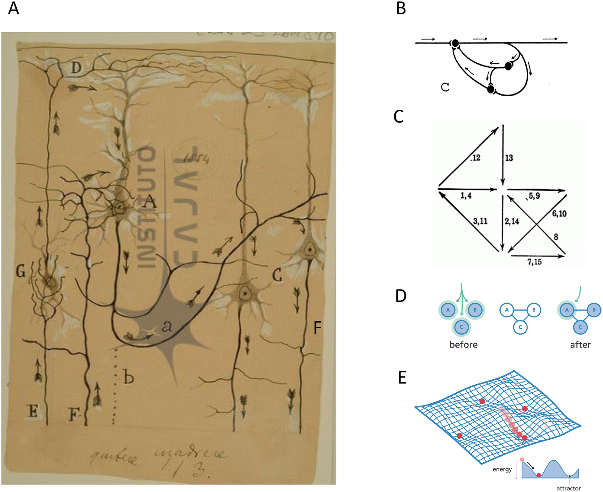
- Ramón y Cajal S (1899). La Textura del Sistema Nerviosa del Hombre y los Vertebrados (Moya; (Primera Edicion)).
-
- Lorente de No R (1938). Analysis of the activity of the chains of internuncial neurons. J. Neurophysiol 1, 207–244.
-
- Hebb DO (1949). The organization of behaviour (Wiley; ).
-
- Semon R (1904). Die Mneme. Engelmann W.
Publication types
MeSH terms
Grants and funding
LinkOut - more resources
Full Text Sources
Research Materials
