. 2024 Jan 23;29(1):18.
doi: 10.1186/s11658-024-00537-1.

Pro-inflammatory cytokines stimulate CFTR-dependent anion secretion in pancreatic ductal epithelium

Affiliations

Pro-inflammatory cytokines stimulate CFTR-dependent anion secretion in pancreatic ductal epithelium

Dora Angyal et al. Cell Mol Biol Lett. .

Abstract

Background: Loss of CFTR-dependent anion and fluid secretion in the ducts of the exocrine pancreas is thought to contribute to the development of pancreatitis, but little is known about the impact of inflammation on ductal CFTR function. Here we used adult stem cell-derived cell cultures (organoids) obtained from porcine pancreas to evaluate the effects of pro-inflammatory cytokines on CFTR function.

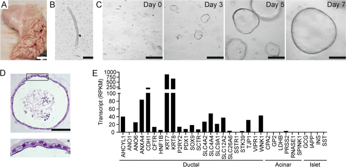
Methods: Organoids were cultured from porcine pancreas and used to prepare ductal epithelial monolayers. Monolayers were characterized by immunocytochemistry. Epithelial bicarbonate and chloride secretion, and the effect of IL-1β, IL-6, IFN-γ, and TNF-α on CFTR function was assessed by electrophysiology.

Results: Immunolocalization of ductal markers, including CFTR, keratin 7, and zonula occludens 1, demonstrated that organoid-derived cells formed a highly polarized epithelium. Stimulation by secretin or VIP triggered CFTR-dependent anion secretion across epithelial monolayers, whereas purinergic receptor stimulation by UTP, elicited CFTR-independent anion secretion. Most of the anion secretory response was attributable to bicarbonate transport. The combination of IL-1β, IL-6, IFN-γ, and TNF-α markedly enhanced CFTR expression and anion secretion across ductal epithelial monolayers, whereas these cytokines had little effect when tested separately. Although TNF-α triggered apoptotic signaling, epithelial barrier function was not significantly affected by cytokine exposure.

Conclusions: Pro-inflammatory cytokines enhance CFTR-dependent anion secretion across pancreatic ductal epithelium. We propose that up-regulation of CFTR in the early stages of the inflammatory response, may serve to promote the removal of pathogenic stimuli from the ductal tree, and limit tissue injury.

Keywords: CFTR; Cystic fibrosis; Cytokines; Epithelial ion transport; Organoid; Pancreatitis.


Conflict of interest statement

The authors declare that no competing interest exists.

Figures

Fig. 1
Fig. 1
Establishment of pancreatic ductal organoids. A Excised pancreatic tissue. Scale bar: 1 cm. B Isolated duct (bright-field microscopy). Scale bar: 100 µm. C Development of spheroid organoids over the first seven days after seeding. (bright-field microscopy). Scale bars: 1 mm. D A pancreatic organoid (HE stain). Scale bar: 100 µm. E Transcript levels of ductal, acinar and islet cell markers
Fig. 2
Fig. 2
Detection of CFTR protein by Western blot analysis in two pancreatic organoid lines (P) and the human intestinal cell line HT29-CL19A (I). Detection of e-cadherin (ECAD) served as a loading control. Outer left lane shows the position of molecular weight markers, with an approximate molecular mass as indicated
Fig. 3
Fig. 3
Pancreatic organoid-derived cells form a polarized epithelium when cultured on a permeable substrate. A Top panel: transverse section of an epithelial monolayer cultured on a Transwell insert (HE-stain). Scale bar: 25 µm. Bottom panel: the apical aspect of a confluent monolayer (bright-field microscopy). Scale bar: 100 µm. B Epithelial monolayers (top) and pancreatic tissue (bottom) were stained for SOX9. Nuclei are visualized using DAPI staining. Insets show SOX9 staining only in the area denoted by an asterisk. Scale bars: 50 µm. C–F Epithelial monolayers (top panels) and pancreatic tissue (bottom panels) were stained for KRT7, ECAD, ZO-1 or CFTR. Nuclei are visualized using DAPI staining. Scale bars: 50 µm. Fluorescence was visualized on a Stellaris 5 low incidence angle upright confocal microscope with a HC PL APO CS2 40x/1.30 oil objective (Leica Microsystems)
Fig. 4
Fig. 4
Pancreatic organoid-derived epithelial monolayers recapitulate ductal anion secretory pathways. A The effect of forskolin (F), secretin (S) and VIP (V) on PKA-mediated phosphorylation of VASP (top panel) and total VASP (bottom panel) levels was assessed by Western blot analysis. Detection of β-actin served as a loading control. Note that phosphorylation of Ser-157 decreases the electrophoretic mobility of VASP. Outer left lane shows the position of molecular weight markers, with an approximate molecular mass as indicated. B Representative experiments showing the effect of secretin, VIP, forskolin and PPQ-102 on the anion secretory current. C The cAMP-dependent Isc responses in nine organoid lines obtained from different animals. Each data point represents one technical replicate. D Forskolin-dependent Isc responses in the presence or absence of bumetanide (Bu). Each data point represents one technical replicate. Data were obtained from two organoid lines. **P= 0.006. Statistical analysis: paired t-test. E Forskolin-dependent Isc responses of monolayers bathed in Meyler solution (M), or in bathing solution containing only bicarbonate as the CFTR-permeating anion. Bicarbonate secretion was assayed at luminal bicarbonate concentrations of 20 or 100 mmol/L, as indicated. Each data point represents one organoid line. F Representative experiment demonstrating the forskolin-dependent Isc response at luminal bicarbonate concentrations of 20 or 100 mmol/L. G Effect of UTP on the anion secretory current. The inset shows the peak UTP-dependent Isc response, assessed in the presence of PPQ-102, in nine organoid lines
Fig. 5
Fig. 5
Effects of pro-inflammatory cytokines on apoptotic signaling and epithelial barrier function in pancreatic ductal organoids and epithelial monolayers. A Transcript levels of cytokine receptors. B Caspase 3/7 activity in epithelial monolayers after 20 h exposure to cytokines. Each data point represents one technical replicate. Statistical analysis: ANOVA. aP = 0.04, bP = 0.02, compared to untreated. C Cleavage of caspase 3 in organoids upon incubation with IL-1β, IL-6, IFN-γ and TNF-α was assessed by Western blot analysis. Detection of β-actin served as a loading control. D FITC-dextran transport across epithelial monolayers in the presence or absence of IL-1β, IL-6, IFN-γ and TNF-α. Data represent the FITC-dextran levels in the contra-luminal compartment, relative to the concentration in the luminal compartment at the start of the experiment. Each data point represents one technical replicate. E Transepithelial electrical resistance (TEER) of epithelial monolayers mounted in Ussing chambers, in the presence or absence of IL-1β, IL-6, IFN-γ and TNF-α. Each data point represents one technical replicate. Statistical analysis: t-test
Fig. 6
Fig. 6
Effect of pro-inflammatory cytokines on anion transport in pancreatic ductal organoid-derived epithelial monolayers. A Effect of combined IL-1β, IL-6, IFN-γ and TNF-α exposure (20 h) on the expression of genes involved in ductal anion secretion. B Representative experiment showing the effect of combined IL-1β, IL-6, IFN-γ and TNF-α exposure (20 h) on the forskolin-dependent, PPQ-102-sensitive, Isc response. C Forskolin-dependent Isc responses in cytokine-treated and control monolayers. The inset shows the change in Isc elicited by PPQ-102. D The effect of different cytokines and combinations thereof on the CFTR-mediated Isc response. Experiments were performed on cultures obtained from three animals. Connecting lines indicate paired experiments performed in parallel, on monolayers cultured from the same organoid line. Statistical analysis: paired t-test (A, C), ANOVA (D). aP = 0.02, bP = 0.007, cP < 0.001, compared to untreated

Similar articles

Cited by

References

    1. Saluja A, Dudeja V, Dawra R, Sah RP. Early intra-acinar events in pathogenesis of pancreatitis. Gastroenterology. 2019;156:1979–1993. doi: 10.1053/j.gastro.2019.01.268. - DOI - PubMed
    1. Habtezion A, Gukovskaya AS, Pandol SJ. Acute pancreatitis: a multifaceted set of organelle and cellular interactions. Gastroenterology. 2019;156:1941–1950. doi: 10.1053/j.gastro.2018.11.082. - DOI - PMC - PubMed
    1. Venkatesh K, Glenn H, Delaney A, Andersen CR, Sasson SC. Fire in the belly: a scoping review of the immunopathological mechanisms of acute pancreatitis. Front Immunol. 2022;13:1077414. doi: 10.3389/fimmu.2022.1077414. - DOI - PMC - PubMed
    1. Watanabe T, Kudo M, Strober W. Immunopathogenesis of pancreatitis. Mucosal Immunol. 2017;10:283–298. doi: 10.1038/mi.2016.101. - DOI - PubMed
    1. Ferrero-Andrés A, Panisello-Roselló A, Roselló-Catafau J, Folch-Puy E. NLRP3 inflammasome-mediated inflammation in acute pancreatitis. Int J Mol Sci. 2020;21:5386. doi: 10.3390/ijms21155386. - DOI - PMC - PubMed