

Correlation between multifocal electroretinogram and optical coherence tomography findings with visual acuity after vitrectomy surgery for retinal detachment: an observational study
- PMID: 38263142
- PMCID: PMC10804544
- DOI: 10.1186/s40942-024-00527-7
Correlation between multifocal electroretinogram and optical coherence tomography findings with visual acuity after vitrectomy surgery for retinal detachment: an observational study
Abstract
Background: Despite the marked increase in the anatomical success rates of macula-off rhegmatogenous retinal detachment (RRD) surgery, patients may still complain about unsatisfactory visual outcome. This study aims to correlate the postoperative corrected distance visual acuity (CDVA) with the mf-ERG (multifocal electroretinogram) and OCT (optical coherence tomography) findings following vitrectomy surgery for RRD.
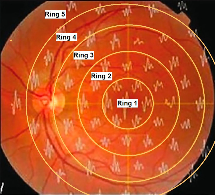
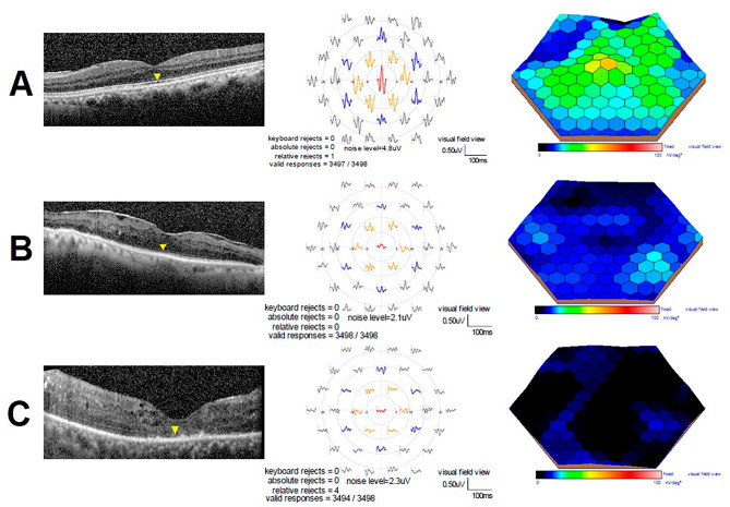
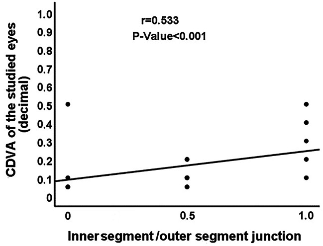
Patients and methods: This retrospective observational study included 40 eyes of 40 patients who underwent successful vitrectomy surgery for macula-off RRD. CDVA, mf-ERG amplitudes, mf-ERG latencies, the central macular thickness (CMT) and the integrity of the inner segment/outer segment (IS/OS) junction assessed by OCT, were evaluated 6 months postoperatively. The correlations between CDVA with mf-ERG amplitudes, mf-ERG latencies, central macular thickness, and IS/OS junction integrity were analyzed.
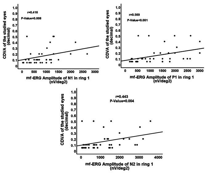
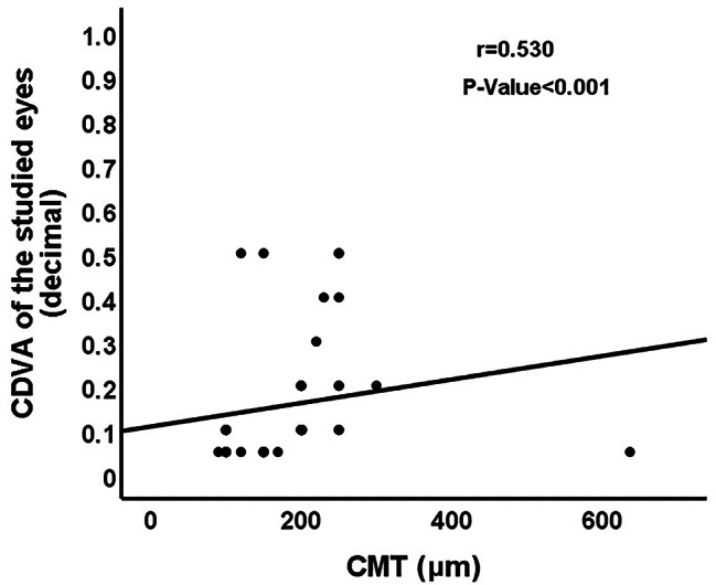
Results: There was a statistically significant moderate positive correlation between CDVA of the studied eyes with mf-ERG amplitudes of N1, P1 and N2 in ring 1 (P = 0.008; P < 0.001 and P = 0.004, respectively), CMT (P < 0.001), and the integrity of IS/OS junction (P < 0.001). There was no significant correlation between CDVA and mf-ERG latencies in ring 1 (P > 0.05). Linear regression analysis revealed that CDVA was significantly associated with mf-ERG amplitudes and the IS/OS junction integrity. In addition, there was a strong positive correlation between mf-ERG amplitudes in ring 1 and the IS/OS junction integrity.
Conclusions: The integrated interpretation of postoperative CDVA, multifocal ERG parameters, and OCT findings provides useful information about functional visual recovery and retinal microstructural changes following vitrectomy for macula-off RRD surgery. The positive correlation between the IS/OS junction integrity and the mf-ERG amplitudes was stronger than the correlation between the IS/OS junction integrity and CDVA suggesting that mf-ERG may be superior to CDVA in reflecting the extent of microstructural damage in the photoreceptor layer.
Trial registration: Clinicaltrials.gov, NCT05993208. Registered 15 August 2023 - Retrospectively registered, https://classic.
Clinicaltrials: gov/ct2/show/NCT05993208 .
Keywords: CDVA; OCT; Retinal detachment; Vitrectomy; mf-ERG.
© 2024. The Author(s).
Conflict of interest statement
The authors declare that they have no competing interests.
Figures
References
Associated data
LinkOut - more resources
Full Text Sources
Medical
