. 2024 Jan 23;15(1):691.
doi: 10.1038/s41467-024-44714-4.

Exosomal secreted SCIMP regulates communication between macrophages and neutrophils in pneumonia

Affiliations

Exosomal secreted SCIMP regulates communication between macrophages and neutrophils in pneumonia

Xiaolei Pei et al. Nat Commun. .

Abstract

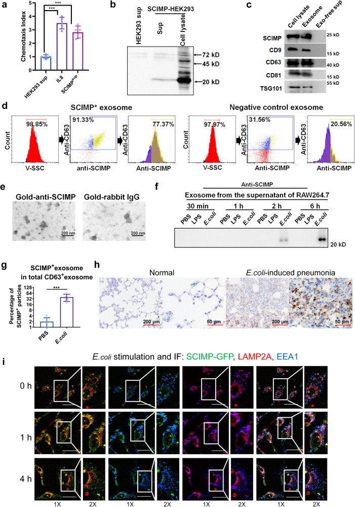
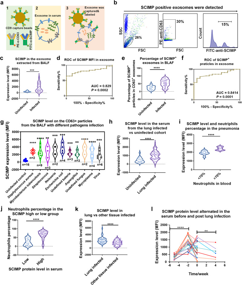
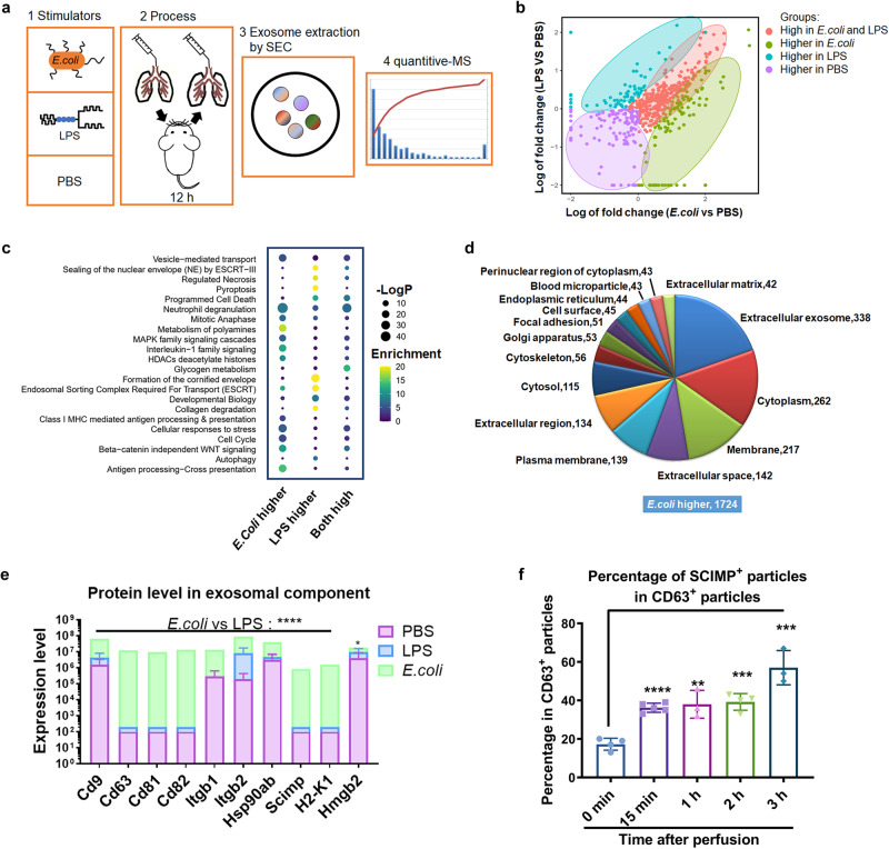
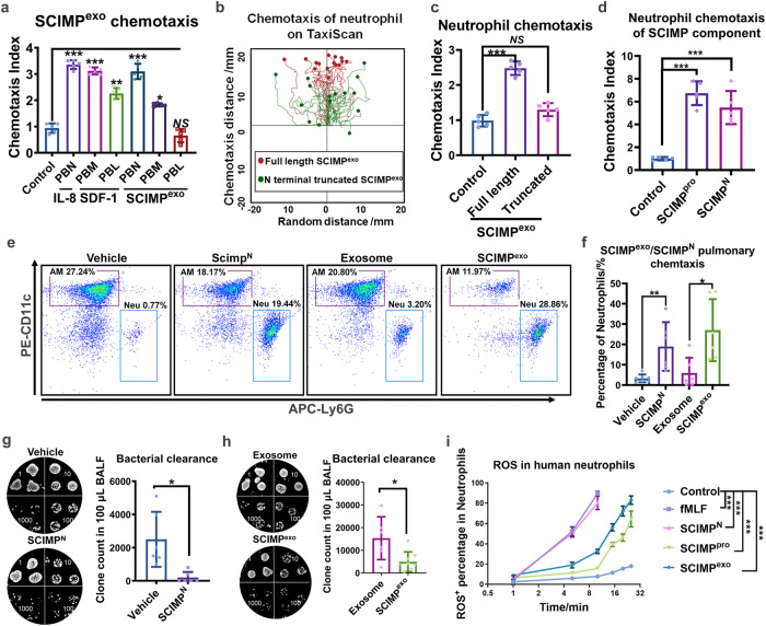
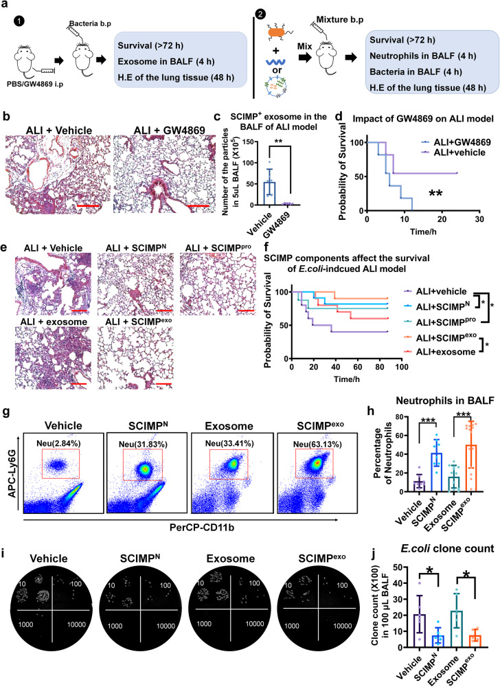
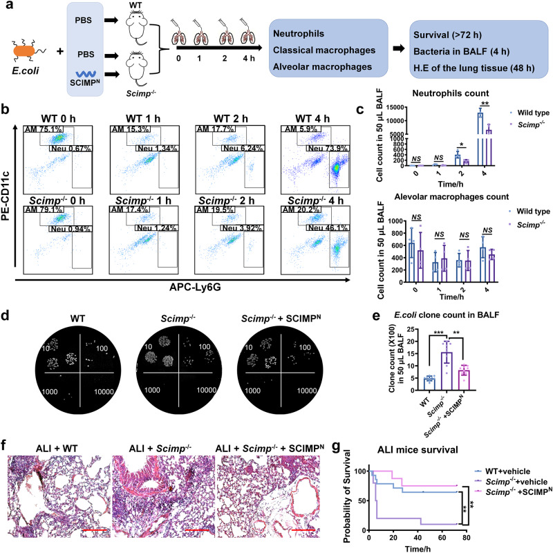
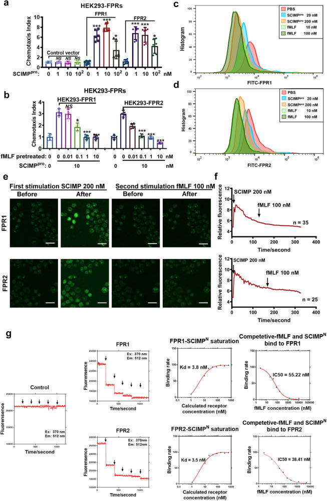
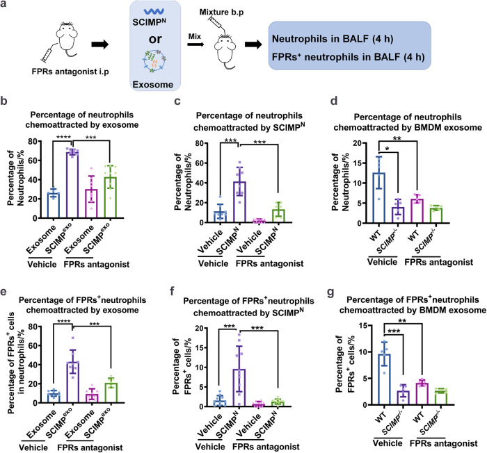
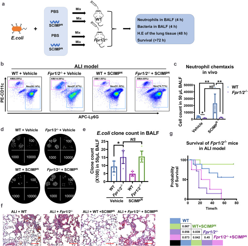
In pneumonia, the deficient or delayed pathogen clearance can lead to pathogen proliferation and subsequent overactive immune responses, inducing acute lung injury (ALI). While screening human genome coding genes using our peripheral blood cell chemotactic platform, we unexpectedly find SLP adaptor and CSK interacting membrane protein (SCIMP), a protein with neutrophil chemotactic activity secreted during ALI. However, the specific role of SCIMP in ALI remains unclear. In this study, we investigate the secretion of SCIMP in exosomes (SCIMPexo) by macrophages after bacterial stimulation, both in vitro and in vivo. We observe a significant increase in the levels of SCIMPexo in bronchoalveolar lavage fluid and serum of pneumonia patients. We also find that bronchial perfusion with SCIMPexo or SCIMP N-terminal peptides increases the survival rate of the ALI model. This occurs due to the chemoattraction and activation of peripheral neutrophils dependent on formyl peptide receptor 1/2 (FPR1/2). Conversely, exosome suppressors and FPR1/2 antagonists decrease the survival rate in the lethal ALI model. Scimp-deficient and Fpr1/2-deficient mice also have lower survival rates and shorter survival times than wild-type mice. However, bronchial perfusion of SCIMP rescues Scimp-deficient mice but not Fpr1/2-deficient mice. Collectively, our findings suggest that the macrophage-SCIMP-FPRs-neutrophil axis plays a vital role in the innate immune process underlying ALI.


Conflict of interest statement

The authors declare no competing interests.

Figures

Fig. 1
Fig. 1. SCIMP can be secreted in exosomes by macrophages stimulated with bacteria.
a The chemotactic activity of the SCIMP-containing supernatant (n = 5) from SCIMP-overexpressing HEK293 cells (SCIMP-HEK293) toward peripheral neutrophils was measured using a chemotaxis assay, with Interleukin 8 (IL-8, 10 ng/mL) used as the positive control (n = 5). b The exogenous SCIMP-6×his recombinant protein in the HEK293 cells could be detected by the anti-6×his antibody both in the cell culture supernatant and cell lysate via western blot. c The exosomes present in the cell culture supernatant of SCIMP-HEK293 were purified using the ultracentrifugation method, and the presence of SCIMP-6×his recombinant protein was confirmed by western blot analysis, along with the exosomal markers CD9, CD63, CD81 and TSG101. d The exosomes from SCIMP-HEK293 or the control HEK293 were analyzed by NTF, and the diameter of the particles was indicated by the V-SSC channel, and PE-anti-CD63 antibody and FITC-anti-SCIMP antibody were used as detection antibodies. e The purified exosomes from SCIMP-HEK293 were observed under the TEM, stained with gold-conjugated anti-SCIMP antibody (scale bar = 200 nm). f The endogenous SCIMP protein secreted in exosomes by RAW264.7 cells with the stimulation of PBS, LPS (1 μg/mL), or heat-denatured E. coli (MOI = 100) for 0.5, 1, 2, and 6 h, was detected by western blot using anti-SCIMP antibody. g The percentage of SCIMP-positive particles in the total exosomes (CD63 positive), which were purified from the supernatant of RAW264.7 cells treated with PBS (n = 8) or E. coli (MOI = 100, n = 10) for 2 h, were stained and measured by NTF. h The endogenous SCIMP protein in the pulmonary tissue of the mice with E. coli-induced pneumonia was detected by IHC using anti-SCIMP antibody (scale bar = 200 μm/50 μm). i The colocalization of SCIMP (exogenously expressed SCIMP-GFP recombinant protein, green), EEA1 (stained with anti-EEA1 antibody, blue), and LAMP2A (stained with anti-LAMP2A antibody, red) was observed by confocal microscopy (IF, Immunofluorescence) in RAW264.7 cells stimulated with heat-denatured E. coli (MOI = 1000) for 0, 1, and 4 h (scale bar = 20 μm). The raw data is available in the “Source Data”.
Fig. 2
Fig. 2. The exosomal SCIMP protein in the pneumonia-related patients was detected using the SCIMPexo detection kit.
a The SCIMP detection kit is schematically shown. b The gating strategy for detecting SCIMP-positive exosomes is shown. cf The relative expression level (c) and the percentage of SCIMP+ particles in CD63+ particles (e) in BALF samples from patients with pulmonary infection (n = 36) or without pulmonary infection (n = 20) were measured using the SCIMPexo detection kit; The ROC curves were generated based on the exosomal SCIMP expression level (d, p = false positive rate) or SCIMP+ particles percentage (f, p = false positive rate) in the pulmonary-infected (n = 36) and -uninfected groups (n = 20), and the AUC was calculated. g The expression level of exosomal SCIMP in CD63+ exosome in BALF samples from pulmonary-infected patients was statistically compared according to the pathogen types. h The exosomal SCIMP expression level of the serum samples from the pulmonary-infected (n = 57)/uninfected (n = 50) patients were measured and statistically compared. i The serum samples from the pulmonary-infected patients were subgrouped based on the neutrophil percentage in the white blood cells (>70%, n = 30 or <70%, n = 27), and the exosomal SCIMP expression level of the serum samples in these two subgroups was measured and analyzed. j The serum samples from the pulmonary-infected patients were subgrouped according to the exosomal SCIMP expression level in the serum (>2000, n = 40 or <2000, n = 17) and the neutrophils percentage in the white blood cells in these two subgroups was measured and analyzed. k The exosomal SCIMP expression level of the serum samples from the patients with pulmonary infection (n = 57) and those with other tissue infections (n = 40) was measured and compared. l The exosomal SCIMP expression level of serum samples collected at different time points from pulmonary-infected patients (n = 34) were measured, with time 0 being when the patient was first diagnosed with infectious fever. The raw data is available in the “Source Data”.
Fig. 3
Fig. 3. In the ALI models, E. coli induced more exosomes to be released into the BALF than LPS.
a The schematic workflow for proteomic analysis of the exosomes extracted from the BALF in the ALI models using different stimulators. b According to the MS results, the proteins distribution in the two-dimensional plot was based on the fold change of the LPS group versus PBS group and the E. coli group versus PBS group; The proteins were classified into four subgroups including high protein level in the E. coli-treated subgroup (green), high protein level in the LPS-treated subgroup (blue), high protein levels in the PBS-treated subgroup (purple), and high protein levels in both the E. coli-treated subgroup and LPS-treated subgroup (red). c The enriched functions were analyzed (P is estimating significance) and shown based on the alteration of protein level in three different treatment groups. d The proportion of the subcellular localizations enriched by the proteins in the subgroup with higher expression in the E. coli-treated mice that those in LPS-treated mice was calculated. e The level of exosomal proteins, including SCIMP, in the BALF of the three groups were summarized (n = 3). f In the E. coli-induced ALI model, BALF was collected at 0 min, 15 min, 1 h, 2 h, and 3 h after E. coli perfusion (n = 4), and the percentage of SCIMP-positive particles was measured using the SCIMPexo detection kit. The raw data is available in the “Source Data”.
Fig. 4
Fig. 4. SCIMP can chemoattract the peripheral neutrophils both in vitro and in vivo.
a After separating human peripheral blood neutrophils (PBN), monocytes (PBM), and lymphocytes (PBL), the chemotactic activity of 1 nM IL-8, 1 nM SDF-1, and 1 mg/mL SCIMP-positive exosomes (SCIMPexo) to these cells in vitro were measured using the TaxiScan system (n = 5). b, c The chemotaxis ability of the full-length SCIMP+ exosomes (1 mg/mL) and the extramembrane N-terminus truncated SCIMP+ exosomes (1 mg/mL) to human peripheral blood neutrophils in vitro were measured using the TaxiScan system (n = 5); SCIMP- exosomes (1 mg/mL) acted as the control in (c). d The chemotactic activity of the purified full-length SCIMP protein (1 nM) and the SCIMPN (1 nM) to human peripheral blood neutrophils in vitro were measured using the TaxiScan system (n = 5). e, f The pulmonary in situ chemotaxis activity of the murine SCIMPN (10 μg in 50 μL PBS per mouse), vehicle buffer (PBS, 50 μL), murine SCIMPexo (10 mg in 50 μL PBS per mouse), and the control exosomes, purified from CHO cells (10 mg in 50 μL PBS per mouse), was determined by measuring the percentage of neutrophils (Neu) in the BALF at 4 h post bronchial perfusion of these stimulators (n = 6). g, h After the mice were pretreated the mice with murine SCIMPN (10 μg in 50 μL PBS per mouse), SCIMP-negative exosomes (10 mg in 50 μL PBS per mouse), or SCIMP-positive exosomes (10 mg in 50 μL PBS per mouse) via b.p for 2 h, the bacteria (E. coli) count in the BALF was measured at 4 h after the bacteria bronchial perfusion (1 × 106 CFU per mouse, n = 6 in each group). i The activity of fMLF (1 nM), SCIMPN (1 nM), SCIMP protein (1 nM), and SCIMPexo (1 mg/mL) on the ROS release of human peripheral blood neutrophils was detected using cytometry at the time points of 1, 5, 10, 15, 20, 25 min after the stimulation (n = 2). The raw data is available in the “Source Data”.
Fig. 5
Fig. 5. SCIMP can elevate the survival rate of the E. coli-induced ALI model by chemoattracting more neutrophils to lung.
a Two schematic workflows show the processes to explore the effect of exosome inhibitor on the ALI model and the effect of murine SCIMPN/SCIMP+ exosome on the ALI model (i.p: intraperitoneal injection; b.p: bronchial perfusion). bd After C57 mice were pretreated with the exosome inhibitor GW4869 (50 μg per mouse) or vehicle for 1 h, E. coli (1 × 107 CFU per mouse) was perfused into the lung; The pulmonary tissue injury stained with H.E (bar = 200 μm), the SCIMP+ exosomes in the BALF, and the survival rate of the ALI model were detected (n = 6 in each group). e, f Mice were perfused with the E. coli (1 × 107 CFU per mouse) accompanied by murine SCIMPN (10 μg per mouse), murine full-length SCIMPpro (100 μg per mouse), the vehicle of peptides/protein (PBS, 50 μL per mouse), murine SCIMP+ exosome (10 mg in 50 μL PBS per mouse), or SCIMP- exosomes (10 mg in 50 μL PBS per mouse), and the effect of SCIMPN, SCIMPpro, and SCIMPexo on the lung injury and survival rate of ALI model were evaluated (bar = 200 μm, n = 10 in each group). g, h Neutrophils (CD11b+Ly6G+F4/80-) in the BALF of E. coli (1 × 107 CFU per mouse) induced ALI model (n = 8 in each group) treated with the vehicle buffer (PBS, 50 μL per mouse), SCIMPN (50 μg per mouse), SCIMP- exosomes from CHO cells (10 mg in 50 μL PBS per mouse), or murine SCIMP+ exosomes (10 mg in 50 μL PBS per mouse), respectively, at 4 h of the model were detected by cytometry. i, j The E. coli clone counts in the BALF of E. coli-induced ALI model (n = 6 in each group) treated with the vehicle buffer (PBS, 50 μL per mouse), SCIMPN (50 μg per mouse), SCIMP-negative exosomes from CHO cells (10 mg in 50 μL PBS per mouse), and murine SCIMP+ exosomes (10 mg in 50 μL PBS per mouse), respectively, at 4 h of the model were determined. The raw data is available in the “Source Data”.
Fig. 6
Fig. 6. SCIMP plays a key role in neutrophil chemotaxis, bacterial clearance, and survival in the ALI model.
a The schematic workflow shows the study of immune response and survival of the ALI model in WT and Scimp-/- mice. After perfusing WT and Scimp-/- mice with E. coli suspension (5 × 106 CFU per mouse) respectively along with the murine SCIMPN (50 μg per mouse) or the vehicle buffer (PBS, 50 μL per mouse), the survival rates of the three groups were observed and analyzed. b, c The count of neutrophil (Neu, CD11b+Ly6G+F4/80-) and alveolar macrophage (AM, CD11b+CD11c+F4/80+) in the BALF of E. coli-induced ALI model in WT mice (n = 6) and Scimp-/- mice (n = 6) were measured by cytometry at 0, 1, 2, and 4 h (two-sided t-test). d, e The bacterial clone count at 4 h of E. coli-induced ALI model in WT mice (n = 6) and Scimp-/- mice (n = 6) with or without rescued by murine SCIMPN (50 μg per mouse) were determined and statistically calculated by diluted bacterial culture. f, g The pulmonary tissue injury and inflammatory cell infiltration observed under the H.E staining biopsies (bar = 200 μm) and the survival of E. coli-induced ALI model of the WT and Scimp-/- mice with or without rescued by bronchus-perfused murine SCIMPN (50 μg per mouse) were evaluated. The raw data is available in the “Source Data”.
Fig. 7
Fig. 7. The FPR1/2 are identified as the receptors of SCIMP with a high affinity.
a The chemotactic activity of SCIMPpro (0, 1, 10, 100 nM) to FPR1/2 was determined by the Boyden Chamber chemotactic assay using the FPR1/2-HEK293 cells or the HEK293 cells transfected with the empty plasmid (n = 5 in each group). b After preincubating the cells with fMLF at different concentrations (0, 0.01, 0.1, 1, 10 nM) to desensitize the FPRs for 1 h, the chemotaxis activity of SCIMPpro (10 nM) to HEK293 cell lines expressing FPR1 or FPR2 was measured by the Boyden Chamber chemotaxis assay (n = 6 in each group). c, d After incubating FPR1/2-HEK293 cells with fMLF (10, 100 nM) and SCIMPpro (20, 200 nM) for 30 min, the distribution of FPR1/FPR2 on the cell membrane of these HEK293 cells was detected by cytometry with FITC-anti-FPR antibodies. e, f After FPR1/2-HEK293 cells were loaded with the Ca2+ probe, the time-coursed calcium influx in the cells stimulated by SCIMPpro (200 nM) for 180 s and then stimulated by fMLF (100 nM) subsequently was recorded under the confocal microscopy (scale bar = 50 μm). g After labeled with BODIPY FL Iodoacetamide, the SCIMPN (final concentration: 1, 10, 20, 40, 80, 160, 320, 640 nM) were incubated with a fixed amount of membrane component of FPR1/2-HEK293 cells (total protein amount: 100 mg per well), and the fluorescence density at the emission wavelength (512 nm) was measured, as well as the fMLF (5, 10, 20, 40, 80, 160, 320, 640, 1280 nM) to competitive binding to the dye labeled SCIMPN (10 μM) and FPR1/2 (total protein amount: 100 mg per well). The raw data is available in the “Source Data”.
Fig. 8
Fig. 8. The FPRs antagonist could inhibit the chemotaxis of SCIMP to neutrophils in vivo.
a The schematic workflow shows that pretreatment with FPRs antagonist (CsH, 50 μg per mouse) for 1 h can inhibit the chemotaxis of bronchus-perfused murine SCIMPN (50 μg per mouse), exogenous murine SCIMPexo (10 mg per mouse), and endogenous murine exosomes (10 mg per mouse) to neutrophils in the pulmonary situ. b, e The chemotaxis activity of murine SCIMPexo (10 mg per mouse) from the supernatant of SCIMP-overexpressing CHO cells and the control exosomes (from the supernatant of empty vector transfected CHO cells, 10 mg per mouse) to neutrophils or FPRs+ neutrophils in the lung of C57 mice was assessed with or without pretreatment of FPRs antagonist using flow cytometry (n = 6 in each group). c, f The chemotaxis activity of the murine SCIMPN (50 μg in 50 μL per mouse) and the vehicle (PBS, 50 μL per mouse) to neutrophils or FPRs+ neutrophils in the lung of C57 mice with or without pretreatment of FPRs antagonist was measured by flow cytometry (n = 9 in each group). d, g The chemotaxis activity of the ultracentrifuge purified endogenous exosomes released by WT or Scimp-/- BMDMs to neutrophils or FPRs+ neutrophils in the lung of C57 mice with or without pretreatment with FPRs antagonist was measured by flow cytometry (n = 5 in each group). The raw data is available in the “Source Data”.
Fig. 9
Fig. 9. FPRs expressed on neutrophils are necessary for the effect of SCIMP on the ALI model.
a The schematic workflow shows the study of the effect of murine SCIMPN on the ALI model by using WT or Fpr1/2-/- mice. bg After transbronchially perfused with SCIMPN (50 μg/50 μL PBS per mouse mixed with 5 × 106 CFU E. coli, n = 5) or Vehicle (50 μL PBS per mouse mixed with 5 × 106 CFU E. coli, n = 5) for 4 h to WT or Fpr1/2-/- mice, neutrophils (Neu, CD11b+CD11c-Ly6G+) and alveolar macrophages (AM, CD11b+CD11c+F4/80+Ly6G-) in the BALF from the ALI model were measured by flow cytometry (b, c), and the bacterial count was measured by dilutional bacterial clone culture (d, e, n = 5), and the lung injury and inflammation were evaluated by histological examination of lung biopsies stained with H.E. (bar = 200 μm, f), and the survival of the four groups (n = 8 in each group) was observed and analyzed (g). The raw data is available in the “Source Data”.

Similar articles

Cited by

References

    1. Draber P, et al. SCIMP, a transmembrane adaptor protein involved in major histocompatibility complex class II signaling. Mol. Cell. Biol. 2011;31:4550–4562. doi: 10.1128/MCB.05817-11. - DOI - PMC - PubMed
    1. Luo L, et al. SCIMP is a transmembrane non-TIR TLR adaptor that promotes proinflammatory cytokine production from macrophages. Nat. Commun. 2017;8:14133. doi: 10.1038/ncomms14133. - DOI - PMC - PubMed
    1. Lucas RM, et al. SCIMP is a spatiotemporal transmembrane scaffold for Erk1/2 to direct pro-inflammatory signaling in TLR-activated macrophages. Cell Rep. 2021;36:109662. doi: 10.1016/j.celrep.2021.109662. - DOI - PubMed
    1. Channappanavar R, et al. Dysregulated type I interferon and inflammatory monocyte-macrophage responses cause lethal pneumonia in SARS-CoV-infected mice. Cell Host Microbe. 2016;19:181–193. doi: 10.1016/j.chom.2016.01.007. - DOI - PMC - PubMed
    1. Yamamoto K, et al. Roles of lung epithelium in neutrophil recruitment during pneumococcal pneumonia. Am. J. Respir. Cell. Mol. Biol. 2014;50:253–262. doi: 10.1165/rcmb.2013-0114OC. - DOI - PMC - PubMed

Substances

LinkOut - more resources