. 2024 Jan 23;24(1):17.
doi: 10.1186/s40644-024-00661-3.

Dual-modal radiomics nomogram based on contrast-enhanced ultrasound to improve differential diagnostic accuracy and reduce unnecessary biopsy rate in ACR TI-RADS 4-5 thyroid nodules

Affiliations

Dual-modal radiomics nomogram based on contrast-enhanced ultrasound to improve differential diagnostic accuracy and reduce unnecessary biopsy rate in ACR TI-RADS 4-5 thyroid nodules

Jia-Yu Ren et al. Cancer Imaging. .

Abstract

Background: American College of Radiology (ACR) Thyroid Imaging Reporting and Data System (TI-RADS, TR) 4 and 5 thyroid nodules (TNs) demonstrate much more complicated and overlapping risk characteristics than TR1-3 and have a rather wide range of malignancy possibilities (> 5%), which may cause overdiagnosis or misdiagnosis. This study was designed to establish and validate a dual-modal ultrasound (US) radiomics nomogram integrating B-mode ultrasound (BMUS) and contrast-enhanced ultrasound (CEUS) imaging to improve differential diagnostic accuracy and reduce unnecessary fine needle aspiration biopsy (FNAB) rates in TR 4-5 TNs.

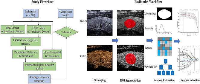
Methods: A retrospective dataset of 312 pathologically confirmed TR4-5 TNs from 269 patients was collected for our study. Data were randomly divided into a training dataset of 219 TNs and a validation dataset of 93 TNs. Radiomics characteristics were derived from the BMUS and CEUS images. After feature reduction, the BMUS and CEUS radiomics scores (Rad-score) were built. A multivariate logistic regression analysis was conducted incorporating both Rad-scores and clinical/US data, and a radiomics nomogram was subsequently developed. The performance of the radiomics nomogram was evaluated using calibration, discrimination, and clinical usefulness, and the unnecessary FNAB rate was also calculated.

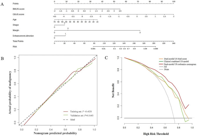
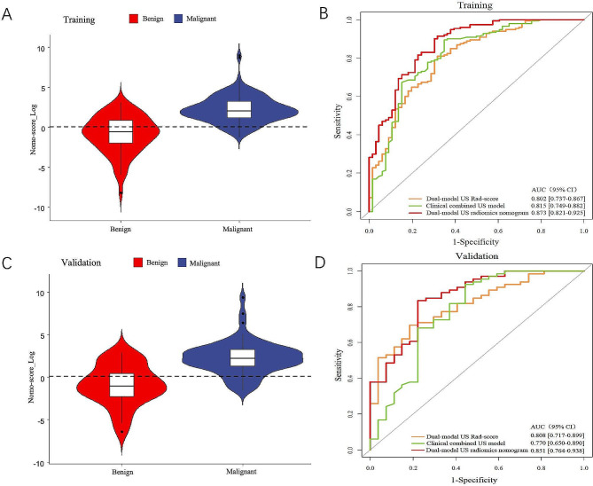
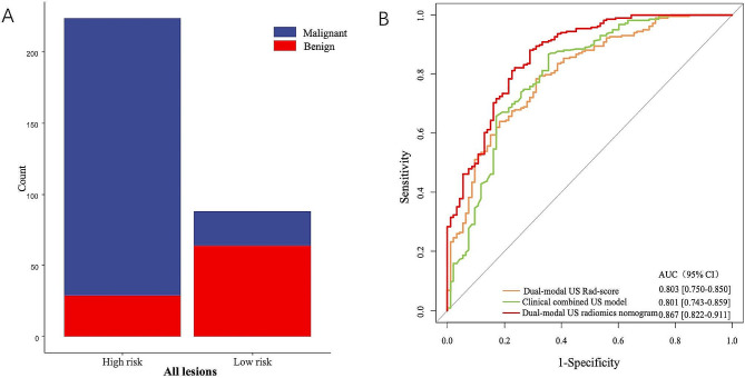
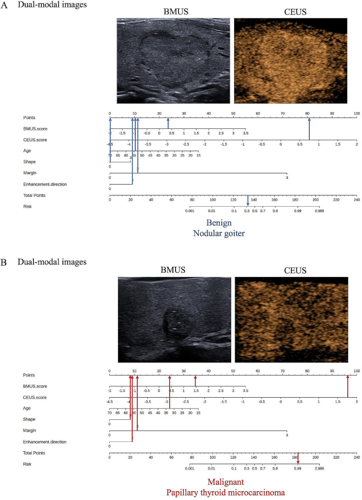
Results: BMUS Rad-score, CEUS Rad-score, age, shape, margin, and enhancement direction were significant independent predictors associated with malignant TR4-5 TNs. The radiomics nomogram involving the six variables exhibited excellent calibration and discrimination in the training and validation cohorts, with an AUC of 0.873 (95% CI, 0.821-0.925) and 0.851 (95% CI, 0.764-0.938), respectively. The marked improvements in the net reclassification index and integrated discriminatory improvement suggested that the BMUS and CEUS Rad-scores could be valuable indicators for distinguishing benign from malignant TR4-5 TNs. Decision curve analysis demonstrated that our developed radiomics nomogram was an instrumental tool for clinical decision-making. Using the radiomics nomogram, the unnecessary FNAB rate decreased from 35.3 to 14.5% in the training cohort and from 41.5 to 17.7% in the validation cohorts compared with ACR TI-RADS.

Conclusion: The dual-modal US radiomics nomogram revealed superior discrimination accuracy and considerably decreased unnecessary FNAB rates in benign and malignant TR4-5 TNs. It could guide further examination or treatment options.

Keywords: ACR TI-RADS 4–5; Contrast-enhanced ultrasound; Nomogram; Radiomics; Thyroid nodules.


Conflict of interest statement

The authors declare that they have no competing interests.

Figures

Fig. 1
Fig. 1
The study flowchart and ultrasound radiomics workflow of the present study. BMUS = B-mode ultrasound, CEUS = contrast-enhanced ultrasound, LASSO = least absolute shrinkage and selection operator, Rad-score = radiomics score, US = ultrasound, ROI = region of interest
Fig. 2
Fig. 2
Dual-modal US radiomics nomogram and its predictive performance for TI-RADS 4 and 5 thyroid nodules. (A) A dual-modal US radiomics nomogram was constructed with BMUS Rad-score, CEUS Rad-score, age, shape, margin, and enhancement direction for predicting malignancy of TI-RADS 4–5 thyroid nodules. (B) Calibration curves of the dual-modal US radiomics nomogram in the training and validation cohorts. The red and green lines represent the actual predictive probabilities of malignancy of the nomogram in the training and validation cohorts, respectively, and the dashed black line represents an ideal prediction. (C) A decision curve analysis (DCA) shows the role of three different models in predicting benign and malignant TI-RADS 4–5 thyroid nodules derived from the entire cohort (n = 312). The DCA shows that using the dual-modal US radiomics nomogram (red curve) to predict benign and malignant TI-RADS 4–5 thyroid nodules provided a greater benefit than the clinical combined US model (green curve) and dual-modal US Rad-score (orange curve). BMUS = B-mode ultrasound, CEUS = contrast-enhanced ultrasound, US = ultrasound, Rad-score = radiomics score
Fig. 3
Fig. 3
Differential diagnostic accuracy of dual-modal US radiomics nomogram for TI-RADS 4 and 5 thyroid nodules. The violin plot shows that the dual-modal US radiomics nomogram performed well in predicting benign and malignant TI-RADS 4–5 thyroid nodules in both the training (A) and validation (C) cohorts. The receiver operating characteristic curves of the dual-modal US radiomics nomogram, clinical combined US model, and the dual-modal US Rad-score model are displayed in the training (B) and validation (D) cohorts, respectively. US = ultrasound, Rad-score = radiomics score, AUC = the area under the receiver operating characteristic curve, CI = confidence interval
Fig. 4
Fig. 4
Performance of dual-modal US radiomics nomogram in all 312 TI-RADS 4 and 5 thyroid nodules. (A) The risk-classification performance of the dual-modal US radiomics nomogram. (B) The ROC curve analyses of the three different models. US = ultrasound, Rad-score = radiomics score, AUC = the area under the receiver operating characteristic curve, CI = confidence interval
Fig. 5
Fig. 5
Two illustrative examples to present the clinical utilization of the nomogram as diagrams. (A) The blue arrows demonstrated that a 54-year-old patient (point: 10.25) has a thyroid nodule which has an aspect ratio < 1 (point: 0), lobulated margin (point: 11.25), centripetal enhancement direction (point: 9), BMUS radiomics score of 0.354 (point: 23), and CEUS radiomics score of 0.715 (point: 80.5). This thyroid nodule got a total point of 134, corresponding to the malignancy probability (defined as Nomo-score) of 0.339. Therefore, this thyroid nodule was predicted as benign by the nomogram according to the optimal cutoff value of 0.524 and was eventually pathologically confirmed as a nodular goiter. (B) The red arrows showed that a 33-year-old (point: 24) patient has a thyroid nodule which has an aspect ratio > 1 (point: 8), irregular margin (point: 11), centripetal enhancement direction (point: 9), BMUS radiomics score of 1.468 (point: 34.75), and CEUS radiomics score of 1.752 (point: 96.25). This thyroid nodule got a total point of 183, referring to a Nomo-score of 0. 991. The nomogram eventually produced an accurate result consistent with the pathology outcome of papillary thyroid carcinoma. BMUS = B-mode ultrasound, CEUS = contrast-enhanced ultrasound

Similar articles

Cited by

References

    1. Haugen BR, Alexander EK, Bible KC, Doherty GM, Mandel SJ, Nikiforov YE, et al. 2015 American Thyroid Association Management Guidelines for adult patients with thyroid nodules and differentiated thyroid Cancer: the American Thyroid Association Guidelines Task Force on thyroid nodules and differentiated thyroid Cancer. Thyroid. 2016;26:1–133. doi: 10.1089/thy.2015.0020. - DOI - PMC - PubMed
    1. Haymart MR, Banerjee M, Reyes-Gastelum D, Caoili E, Norton EC. Thyroid ultrasound and the increase in diagnosis of low-risk thyroid Cancer. J Clin Endocrinol Metab. 2019;104:785–92. doi: 10.1210/jc.2018-01933. - DOI - PMC - PubMed
    1. Tessler FN, Middleton WD, Grant EG, Hoang JK, Berland LL, Teefey SA, et al. ACR thyroid imaging, reporting and Data System (TI-RADS): White Paper of the ACR TI-RADS Committee. J Am Coll Radiol. 2017;14:587–95. doi: 10.1016/j.jacr.2017.01.046. - DOI - PubMed
    1. Wang J, Jiang J, Zhang D, Zhang YZ, Guo L, Jiang Y, et al. An integrated AI model to improve diagnostic accuracy of ultrasound and output known risk features in suspicious thyroid nodules. Eur Radiol. 2022;32:2120–9. doi: 10.1007/s00330-021-08298-7. - DOI - PubMed
    1. Radzina M, Ratniece M, Putrins DS, Saule L, Cantisani V. Performance of contrast-enhanced ultrasound in thyroid nodules: review of current state and future perspectives. Cancers (Basel). 2021;13. - PMC - PubMed

LinkOut - more resources