. 2024 Jan 23;10(1):10.
doi: 10.1038/s41421-023-00639-8.

Pharmacology of LRRK2 with type I and II kinase inhibitors revealed by cryo-EM

Affiliations

Pharmacology of LRRK2 with type I and II kinase inhibitors revealed by cryo-EM

Hanwen Zhu et al. Cell Discov. .

Erratum in

Abstract

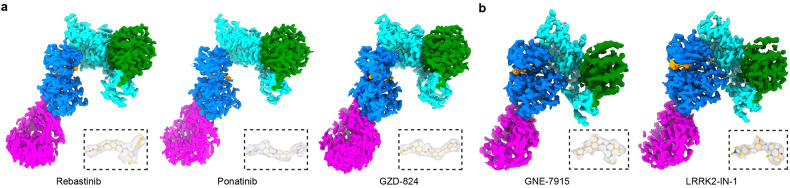
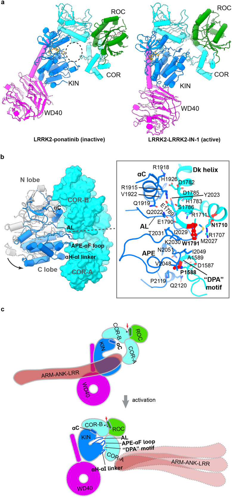
LRRK2 is one of the most promising drug targets for Parkinson's disease. Though type I kinase inhibitors of LRRK2 are under clinical trials, alternative strategies like type II inhibitors are being actively pursued due to the potential undesired effects of type I inhibitors. Currently, a robust method for LRRK2-inhibitor structure determination to guide structure-based drug discovery is lacking, and inhibition mechanisms of available compounds are also unclear. Here we present near-atomic-resolution structures of LRRK2 with type I (LRRK2-IN-1 and GNE-7915) and type II (rebastinib, ponatinib, and GZD-824) inhibitors, uncovering the structural basis of LRRK2 inhibition and conformational plasticity of the kinase domain with molecular dynamics (MD) simulations. Type I and II inhibitors bind to LRRK2 in active-like and inactive conformations, so LRRK2-inhibitor complexes further reveal general structural features associated with LRRK2 activation. Our study provides atomic details of LRRK2-inhibitor interactions and a framework for understanding LRRK2 activation and for rational drug design.


Conflict of interest statement

The authors declare no competing interests.

Figures

Fig. 1
Fig. 1. Overall structures of LRRK2RCKW bound to type I or type II inhibitors.
a Cryo-EM maps of LRRK2RCKW bound to type II inhibitors. Left: LRRK2–rebastinib complex; Middle: LRRK2–ponatinib complex; Right: LRRK2–GZD-824 complex. Only C-terminal catalytic halves of LRRK2 (LRRK2RCKW) bound to type II inhibitors are shown for simplicity and better comparison. b Cryo-EM maps of LRRK2 bound to type I inhibitors. Left: LRRK2RCKWm–GNE-7915 complex; Right: LRRK2RCKWm–LRRK2-IN-1 complex. The cryo-EM maps are colored by domain architecture: ROC green, COR cyan, KIN blue, WD40 magenta, inhibitor yellow. Cryo-EM density maps of inhibitors are shown in dashed boxes.
Fig. 2
Fig. 2. Binding of type II inhibitors to the LRRK2 KIN domain.
ac Interactions between rebastinib (a), ponatinib (b), or GZD-824 (c) and the LRRK2 KIN domain. Left: stereo view of the inhibitor-binding site; Right: schematic drawing of interactions formed between inhibitors and LRRK2. The bound inhibitors in yellow and surrounding residues involved in the binding are shown as ball sticks and labeled. Dashed lines indicate hydrophilic interactions. d Interactions between ATP and the inactive LRRK2 KIN domain (PDB 7LI4). Left: stereo view of the ATP-binding site; Right: schematic drawing of interactions formed between ATP molecule and LRRK2 KIN domain. e KIN domain comparison between the ATP-bound inactive LRRK2 (gray) and type-II inhibitor-bound LRRK2 structures (LRRK2–rebastinib: red, LRRK2–ponatinib: blue, LRRK2–GZD824: green). Gly loop, activation loop, and αC helix are highlighted and compared. The ATP or inhibitor binding pocket in LRRK2 is indicated by gray and orange dashed circles, respectively. f Comparison of the active site between ATP-bound inactive LRRK2 and the ponatinib-bound LRRK2. Differences between the Gly loop, αC helix, K1906-E1920 salt bridge, Y2018 configuration, and activation loop are indicated. Ponatinib is shown as a yellow surface.
Fig. 3
Fig. 3. Binding of type I inhibitors to the LRRK2 KIN domain.
ac Interactions between GNE-7915 (a), LRRK2-IN-1 (b) or DNL201 (c) (PDB 8SMC) and LRRK2 KIN domain. Left: stereo view of the inhibitor-binding site; Right: schematic drawing of interactions formed between inhibitors and LRRK2. The inhibitors, colored in yellow, and surrounding residues are shown as ball sticks and labeled. Dashed lines indicate hydrophilic interactions. d Interactions between ATP and the active LRRK2 KIN domain (PDB 8FO9). Left: stereo view of the ATP-binding site; Right: schematic drawing of interactions formed between ATP molecule and the LRRK2-binding site.
Fig. 4
Fig. 4. LRRK2 activation mechanism revealed by type I or type II inhibitors.
a Structural comparison between LRRK2–ponatinib (left) and LRRK2–LRRK2-IN-1 (right) complexes. A dashed circle indicates the “central cavity” between the KIN and COR domains. b Movement of the KIN domain relative to the COR domain upon LRRK2-IN-1 binding compared to ponatinib binding (gray). Key structural elements are labeled. Interactions between the KIN and COR domains in the LRRK2 bound to LRRK2-IN-1 are illustrated. Sidechains of interface residues are shown. The residues, shown to be important for LRRK2 active state stabilization, are highlighted in red. c Cartoon representation illustrating common features in LRRK2 activation.
Fig. 5
Fig. 5. MD simulation of LRRK2 KIN domain.
a Metastable states (S0–S4) revealed by GaMD simulations. The free energy profile was projected along two RMSD coordinates (see the section “Materials and methods”). b Comparison between the S0 state and the ATP-bound active state. c Comparison between the S4 state and the ATP-bound inactive state. d LRRK2–ponatinib vs S1–S3 states. e LRRK2–rebastinib vs S1–S3 states. f LRRK2–GZD-824 vs S1–S3 states. Key structural elements, including Gly loop, activation loop (AL), and αC helix, are highlighted with S1–S3 in blue, magenta, and brown, respectively.

Similar articles

Cited by

References

    1. Dorsey ER, Sherer T, Okun MS, Bloem BR. The emerging evidence of the Parkinson pandemic. J. Parkinsons Dis. 2018;8:S3–S8. - PMC - PubMed
    1. Taylor M, Alessi DR. Advances in elucidating the function of leucine-rich repeat protein kinase-2 in normal cells and Parkinson’s disease. Curr. Opin. Cell Biol. 2020;63:102–113. - PMC - PubMed
    1. Usmani A, Shavarebi F, Hiniker A. The cell biology of LRRK2 in Parkinson’s disease. Mol. Cell. Biol. 2021;41:e00660–20. - PMC - PubMed
    1. Alexander Boecker C. The role of LRRK2 in intracellular organelle dynamics. J. Mol. Biol. 2023;435:167998. - PubMed
    1. Bonet-Ponce L, Cookson MR. LRRK2 recruitment, activity, and function in organelles. FEBS J. 2022;289:6871–6890. - PMC - PubMed