. 2024 Jan 23;10(1):9.
doi: 10.1038/s41421-023-00632-1.

Integrated compact regulators of protein activity enable control of signaling pathways and genome-editing in vivo

Affiliations

Integrated compact regulators of protein activity enable control of signaling pathways and genome-editing in vivo

Nik Franko et al. Cell Discov. .

Abstract

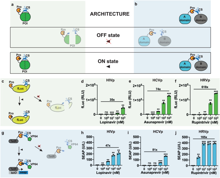
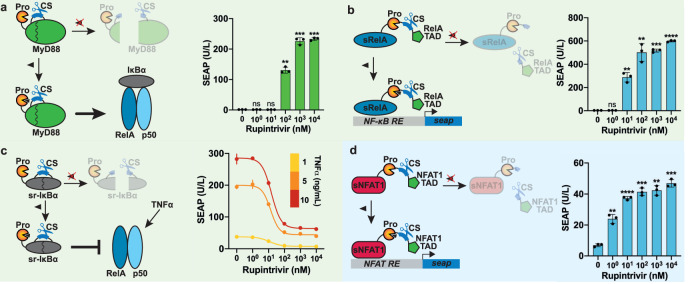
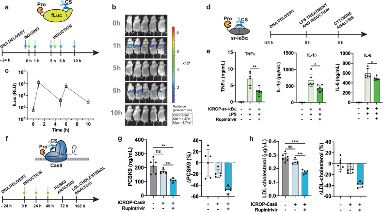
Viral proteases and clinically safe inhibitors were employed to build integrated compact regulators of protein activity (iCROP) for post-translational regulation of functional proteins by tunable proteolytic activity. In the absence of inhibitor, the co-localized/fused protease cleaves a target peptide sequence introduced in an exposed loop of the protein of interest, irreversibly fragmenting the protein structure and destroying its functionality. We selected three proteases and demonstrated the versatility of the iCROP framework by validating it to regulate the functional activity of ten different proteins. iCROP switches can be delivered either as mRNA or DNA, and provide rapid actuation kinetics with large induction ratios, while remaining strongly suppressed in the off state without inhibitor. iCROPs for effectors of the NF-κB and NFAT signaling pathways were assembled and confirmed to enable precise activation/inhibition of downstream events in response to protease inhibitors. In lipopolysaccharide-treated mice, iCROP-sr-IκBα suppressed cytokine release ("cytokine storm") by rescuing the activity of IκBα, which suppresses NF-κB signaling. We also constructed compact inducible CRISPR-(d)Cas9 variants and showed that iCROP-Cas9-mediated knockout of the PCSK9 gene in the liver lowered blood LDL-cholesterol levels in mice. iCROP-based protein switches will facilitate protein-level regulation in basic research and translational applications.


Conflict of interest statement

The authors declare no competing interests.

Figures

Fig. 1
Fig. 1. Compact small-molecule-responsive protein switches.
a, b Schematic representation of the protein switches. The POI is modified to contain a protease cleavage site (CS) in a permissible location, and the corresponding protease is either a fused to the N-terminus or b placed next to the CS, between two domains (A and B) of the POI. In both configurations, the protease inactivates the functionality of the effector protein (OFF state). The addition of a small molecule inhibitor blocks the protease activity, thereby preserving the activity of the effector protein (ON state). c Scheme of synthetic post-translational regulation of fLuc. The protease CS was placed downstream of fLuc residue K491 and the protease from d HIV, e HCV, or f HRV was fused at the N-terminus. df Bioluminescence intensity of HEK293T cells transfected with each fLuc protease system and treated for 24 h with different concentrations of the corresponding inhibitors. g Scheme of synthetic post-translational regulation of the transcription factor TetR-VP64. The protease from h HIV, i HCV, or j HRV and cognate CSs were placed between TetR and VP64. hj SEAP secretion from HEK293T cells co-transfected with each TetR-VP64 protease system and a tetO-driven SEAP reporter, and treated for 24 h with different concentrations of the corresponding inhibitors. In d, e, f, h, i, j data are shown as mean ± SD, with individual data points (n = 3 biological replicates). Numbers above bars indicate fold difference between values at the highest concentration and un-induced cells. Statistical significance was calculated by means of Welch’s two-tailed t-test, *P < 0.05, **P < 0.01, ***P < 0.001, ****P < 0.0001, ns, not significant.
Fig. 2
Fig. 2. Kinetics and reversibility of iCROP-modified fLuc delivered as DNA or mRNA.
a Schematic representation of the post-translational steps that iCROP-fLuc undergoes in the presence or absence of the protease inhibitor. b FLuc luminescence every 15 to 30 min during 90 min of incubation with rupintrivir at the indicated concentrations. HEK293T cells were transfected with iCROP-fLuc (PmPGK-HRVpΔQ182-fLucCS-K491-pA) and 36 h later the medium was exchanged for fresh medium either without or with rupintrivir. c Real-time fLuc analysis from HEK293T cells expressing iCROP-fLuc. The recording of the signal from live cells was started 90 s before changing the medium to fresh medium either without or with rupintrivir (100 nM or 1000 nM). d iCROP-fLuc reversibility. HEK293T cells expressing iCROP-fLuc were alternated between rupintrivir-containing (100 nM) and rupintrivir-free media. Each rupintrivir induction lasted 45 min, followed by 135 min in rupintrivir-free medium. fLuc intensity was measured at the indicated time points. e Inducibility of iCROP-fLuc delivered as mRNA. Luminescence from HEK293T cells transfected with in vitro-transcribed mRNA coding for iCROP-fLuc for 6 h, followed by incubation for additional 6 h in the presence of the indicated rupintrivir concentrations. f Activation kinetics of mRNA-delivered iCROP-fLuc. HEK293T cells simultaneously transfected with iCROP-fLuc mRNA and treated with rupintrivir (1 μM) were analyzed for fLuc intensity at the indicated time points. In bf, data are shown as mean ± SD (n = 3 biological replicates). Statistical significance was calculated by means of Welch’s two-tailed t-test, *P < 0.05, **P < 0.01, ***P < 0.001, ****P < 0.0001, ns, not significant.
Fig. 3
Fig. 3. Regulation of endogenous signaling pathways by iCROP-engineered signaling mediators.
a MyD88-mediated NF-κB activation in response to rupintrivir, which preserves the function of iCROP-engineered MyD88. MyD88 bearing the HRV protease at its N-terminus and the cognate CS at position G83 is fragmented into two inactive parts unless the protease inhibitor is present. When rupintrivir is present, intact MyD88 activates downstream effectors, culminating with the assembly of the complex in the promoter region of NF-κB target genes (left). HEK293T cells co-transfected with constitutive expression of iCROP-MyD88 (PmPGK-HRVpΔQ182-Myd88CS-G83-pA) and SEAP expression from an NF-κB-responsive promoter (PNF-κB-SEAP-pA) were treated with rupintrivir at the indicated concentrations for 24 h before analyzing SEAP expression levels (right). b RelA-mediated NF-κB activation in response to rupintrivir, which preserves the function of iCROP-engineered RelA (left). RelA bearing both the HRVp and CS at position S319 is fragmented into two non-functional parts, unless the protease inhibitor is provided, when transcription of the reporter SEAP can occur. HEK293T cells were co-transfected with constitutively expressed iCROP-RelA (PmPGK-N-RelA-HRVp-RelA-C-pA) and PNF-κB-SEAP-pA and treated with rupintrivir at various concentrations for 24 h before analyzing SEAP expression levels (right). c Super-repressor sr-IκBα-mediated NF-κB repression in response to rupintrivir. sr-IκBα bearing the HRV protease at its N-terminus and the cognate CS at position P170 is fragmented into two inactive parts, unless the protease inhibitor is provided, thereby suppressing NF-κB signaling (left). HEK293T cells co-transfected with iCROP-sr-IκBα and PNFκB-SEAP-pA were challenged with the indicated TNFα concentrations to induce NF-κB signaling and treated with different doses of rupintrivir for 24 h before analyzing SEAP expression levels (right). d NFAT1-mediated activation of NFAT signaling in response to rupintrivir. The HRVp and CS were placed between the truncated DNA-binding domain (sNFAT1S172-G701-DBD) and the transactivation domain (NFAT1L702-T925-TAD) (left). HEK293T cells co-transfected with iCROP-NFAT1 and SEAP expression from an NFAT-responsive promoter (pMX57, P3xNFAT-SEAP-pA) were treated with the indicated rupintrivir concentrations for 24 h before analyzing SEAP expression levels (right). Data are shown as mean ± SD (n = 3 biological replicates) in all panels. Statistical significance was calculated by means of Welch’s two-tailed t-test, *P < 0.05, **P < 0.01, ***P < 0.001, ****P < 0.0001, ns, not significant.
Fig. 4
Fig. 4. Transcriptional regulation and genome editing using iCROP-engineered (d)Cas9 protein or MCPVP64.
a Schematic representation of the iCROP-modified (d)Cas9 protein. dCas9 or Cas9 protein bearing the HRV protease at the N-terminus and the cognate CS at position R535 is fragmented into two inactive parts, unless the protease inhibitor is present. In the presence of inhibitor, (i) active dCas9 associates with a target sgRNA fused to an MS2 loop, which co-localizes the MCP protein fused to a transcriptional activation domain in the promoter region of the target gene, thereby activating transcription; and (ii) active Cas9 associates with a target sgRNA introducing mutations in a target genomic locus. b SEAP produced by cells constitutively expressing iCROP-dCas9 (PmPGK-HRVpΔQ182-dCas9CS-R535-pA), MS2 loop fused to an sgRNA targeting the insulin promoter (PU6-sgRNA(PINS)-MS2), MCP fused to the transactivation domain (pKK44, PhCMV-MCP-p65TA-HSF1TA-pA) and SEAP expression under an insulin-responsive promoter (PINS-SEAP-pA), and treated for 24 h with the indicated concentrations of rupintrivir. c Schematic illustration of the iCROP-MCPVP64 system for dCas9-guided transcription regulation in response to rupintrivir. d Activation of gene expression from the endogenous genomic target with the iCROP-MCPVP64 system. Cells were co-transfected with dCas9, iCROP-MCPVP64 and sgRNA targeting the IL-12B promoter (sgRNA(PIL-12B)-MS2) and treated with the specified rupintrivir concentrations for 48 h before analyzing IL-12 levels in the supernatant by ELISA. e Kinetics of iCROP-MCPVP64-based activated IL-12 expression. Time-course analysis of IL-12 levels in culture supernatants from cells treated either with 1 µM rupintrivir or vehicle. f Mutation rate in the EMX1 gene in cells expressing iCROP-Cas9 and sgRNA(EMX1), and treated with indicated concentrations of rupintrivir for 48 h before NGS analysis. Cells co-transfected with non-modified Cas9 and sgRNA(EMX1) were included as a positive control. g Heat-map representing time- and dose-dependent EMX1 relative indel frequency for iCROP-Cas9. Relative indel frequency was calculated as the ratio between the mutation rate at each specified dose-time point and the maximal mutation rate (n = 3 biological replicates). h Frequency of indel mutations in the genomic loci of VEGFA, TNFRSF1A and ACE2 genes in cells co-transfected with iCROP-Cas9 and each sgRNA targeting gene, and kept for 48 h in the absence or presence of rupintrivir (1 μM) (sgRNA variants with the highest fold induction of mutation rates are represented (see Supplementary Fig. S12g for efficiency of other sgRNA). In panels b, d, e, f, h, data are shown as mean ± SD (n = 3 biological replicates). Statistical significance was calculated by means of Welch’s two-tailed t-test, *P < 0.05, **P < 0.01, ***P < 0.001, ****P < 0.0001, ns, not significant.
Fig. 5
Fig. 5. Activity and functional performance of iCROP systems in vivo.
a Timeline for iCROP-fLuc study in vivo. Mice were hydrodynamically injected with the plasmid encoding iCROP-fLuc 24 h before rupintrivir administration. b Luminescence images were recorded at the indicated time points by the IVIS imaging system (n = 5 mice). c Quantification of the luminescence intensity of images in b. d Timeline for the in vivo study using iCROP-sr-IκBα to repress NF-κB signaling. The mice received the iCROP-sr-IκBα-encoding plasmid by hydrodynamic injection 24 h before the LPS treatment and rupintrivir administration. Blood samples were collected for cytokine analysis 6 h later. e Blood levels of TNFα, IL-1β and IL-6 6 h after the LPS treatment, analyzed by ELISA. Data are shown as mean ± SD, with individual data points (n = 6 mice per experimental group, n = 4 mice for wild-type group). f Timeline for the in vivo study using inducible iCROP-Cas9 for PCSK9 knockout. The DNA vectors expressing iCROP-Cas9 and sgRNA(mPCSK9) were delivered to mice 1 day before the first rupintrivir treatment, which was repeated for 3 consecutive days, before analysis. g Serum PCSK9 levels measured by ELISA (left) in wild-type mice, mice expressing only the iCROP-Cas9 system and mice expressing the iCROP-Cas9 system in combination with rupintrivir treatment. Results are shown as absolute PCSK9 values (left) and relative PCSK9 reduction compared to wild-type mice (right). Relative decrease was calculated by dividing the average PCSK9 levels of experimental groups by the average PCSK9 level of the wild-type group. h LDL-cholesterol levels in mouse serum, measured with an LDL-cholesterol assay kit 168 h after starting the rupintrivir dosing (left) and relative LDL-cholesterol reduction compared to wild-type mice (right). Relative decrease was calculated by dividing the average LDL-cholesterol levels of experimental groups by the average LDL-cholesterol level of the wild-type group. In panels g, h, data are shown as mean ± SD, with individual data points (n = 5–6 mice per group). Statistical significance was calculated by means of Welch’s two-tailed t-test, *P < 0.05, **P < 0.01, ***P < 0.001, ****P < 0.0001, ns, not significant.

Similar articles

Cited by

References

    1. Xie M, Fussenegger M. Designing cell function: assembly of synthetic gene circuits for cell biology applications. Nat. Rev. Mol. Cell Biol. 2018;19:507–525. doi: 10.1038/s41580-018-0024-z. - DOI - PubMed
    1. Khalil AS, et al. A synthetic biology framework for programming eukaryotic transcription functions. Cell. 2012;150:647–658. doi: 10.1016/j.cell.2012.05.045. - DOI - PMC - PubMed
    1. Tigges M, Marquez-Lago TT, Stelling J, Fussenegger M. A tunable synthetic mammalian oscillator. Nature. 2009;457:309–312. doi: 10.1038/nature07616. - DOI - PubMed
    1. Ausländer S, Ausländer D, Müller M, Wieland M, Fussenegger M. Programmable single-cell mammalian biocomputers. Nature. 2012;487:123–127. doi: 10.1038/nature11149. - DOI - PubMed
    1. Gaber R, et al. Designable DNA-binding domains enable construction of logic circuits in mammalian cells. Nat. Chem. Biol. 2014;10:203–208. doi: 10.1038/nchembio.1433. - DOI - PubMed