

Imaging of musculoskeletal tuberculosis
- PMID: 38263840
- PMCID: PMC11027299
- DOI: 10.1093/bjr/tqad019
Imaging of musculoskeletal tuberculosis
Abstract
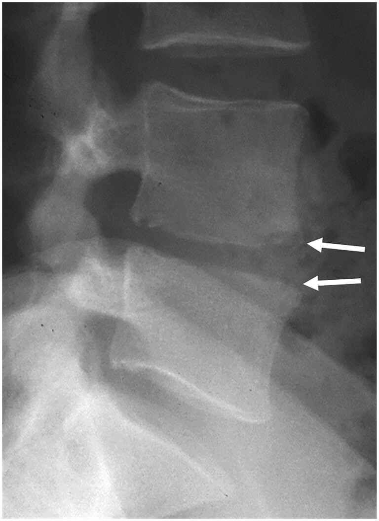
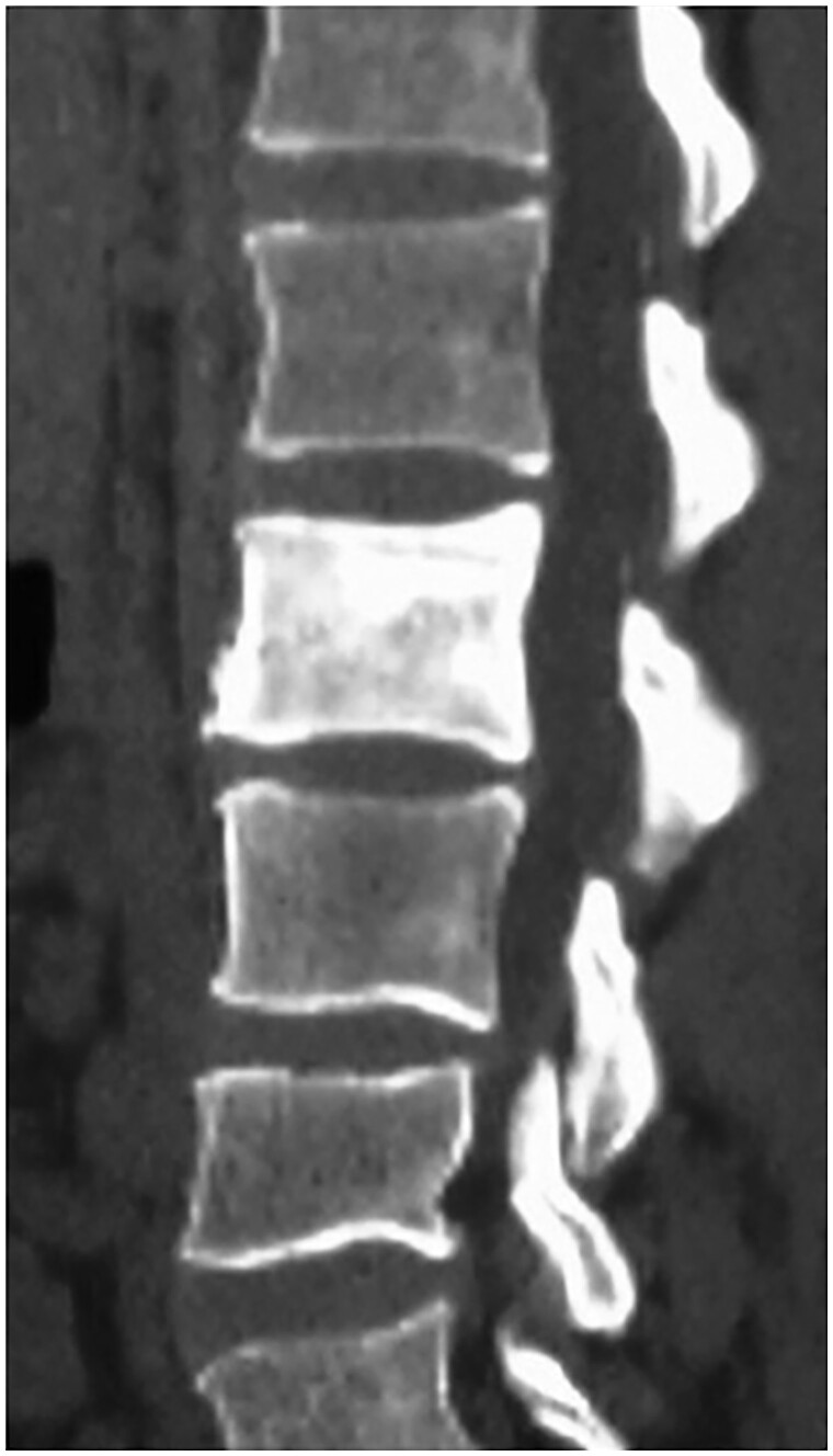
Extra-pulmonary tuberculosis (TB) of the musculoskeletal system usually manifests with non-specific clinical features, mimicking a variety of diseases. Diagnosis and treatment of spinal and extra-spinal musculoskeletal TB are often challenging. Imaging has an important role in detecting this disease, aiding diagnosis, identifying complications, and monitoring disease progression. Radiographs and magnetic resonance imaging are the key imaging modalities utilized. Radiologists should aim to be familiar with the spectrum of imaging features of TB affecting spinal and extra-spinal locations in the musculoskeletal system.
Keywords: bone tuberculosis; extra-pulmonary tuberculosis; joint tuberculosis; musculoskeletal tuberculosis; skeletal tuberculosis; spinal tuberculosis; tuberculosis; tuberculous bursitis; tuberculous osteomyelitis; tuberculous tenosynovitis.
© The Author(s) 2023. Published by Oxford University Press on behalf of the British Institute of Radiology. All rights reserved. For permissions, please email: journals.permissions@oup.com.
Conflict of interest statement
None declared.
Figures


Similar articles
-
Imaging of musculoskeletal tuberculosis.Skeletal Radiol. 2024 Oct;53(10):2081-2097. doi: 10.1007/s00256-023-04556-5. Epub 2024 Jan 17. Skeletal Radiol. 2024. PMID: 38231262 Review.
-
Tuberculosis revisted: classic imaging findings in childhood.Pediatr Radiol. 2023 Aug;53(9):1799-1828. doi: 10.1007/s00247-023-05648-z. Epub 2023 May 23. Pediatr Radiol. 2023. PMID: 37217783 Free PMC article. Review.
-
Atypical extraspinal musculoskeletal tuberculosis in immunocompetent patients: part II, tuberculous myositis, tuberculous bursitis, and tuberculous tenosynovites.Can Assoc Radiol J. 2006 Dec;57(5):278-86. Can Assoc Radiol J. 2006. PMID: 17265982 Review.
-
Tuberculous osteomyelitis and spondylodiscitis.Semin Musculoskelet Radiol. 2011 Nov;15(5):446-58. doi: 10.1055/s-0031-1293491. Epub 2011 Nov 11. Semin Musculoskelet Radiol. 2011. PMID: 22081280 Review.
-
Imaging of musculoskeletal tuberculosis: a new look at an old disease.Clin Orthop Relat Res. 2002 May;(398):32-9. doi: 10.1097/00003086-200205000-00006. Clin Orthop Relat Res. 2002. PMID: 11964629 Review.
Cited by
-
Tuberculous osteo-arthritis unmasked through unusual elbow swelling: A case report.Int J Surg Case Rep. 2025 Jan;126:110759. doi: 10.1016/j.ijscr.2024.110759. Epub 2024 Dec 24. Int J Surg Case Rep. 2025. PMID: 39731801 Free PMC article.
-
Extrapulmonary Comparisons Between Mycobacterium Tuberculosis and Non-Tuberculous Mycobacteria: From Manifestations and Diagnosis to Treatment.Infect Drug Resist. 2025 May 22;18:2613-2627. doi: 10.2147/IDR.S515196. eCollection 2025. Infect Drug Resist. 2025. PMID: 40432812 Free PMC article. Review.
-
A rare case of osteoarticular tuberculosis and tuberculous osteomyelitis of the left foot without pulmonary involvement.Radiol Case Rep. 2024 Sep 28;19(12):6609-6613. doi: 10.1016/j.radcr.2024.09.088. eCollection 2024 Dec. Radiol Case Rep. 2024. PMID: 39380834 Free PMC article.
-
Tuberculosis Beyond the Lungs: A Pictorial Review of Key Diagnostic Imaging Insights.Cureus. 2025 Mar 26;17(3):e81256. doi: 10.7759/cureus.81256. eCollection 2025 Mar. Cureus. 2025. PMID: 40291232 Free PMC article. Review.
-
A Young Man With Tuberculosis in the Neck Soft Tissue: A Case Report.Cureus. 2024 Aug 6;16(8):e66330. doi: 10.7759/cureus.66330. eCollection 2024 Aug. Cureus. 2024. PMID: 39247000 Free PMC article.
References
-
- World Health Organization. Global Tuberculosis Report 2022. Geneva, Switzerland: World Health Organization, 2022. Accessed May 1, 2023. https://www.who.int/publications/i/item/9789240061729
-
- Warner DF, Koch A, Mizrahi V.. Diversity and disease pathogenesis in Mycobacterium tuberculosis. Trends Microbiol. 2015;23(1):14-21. - PubMed
-
- Pai M, Behr MA, Dowdy D, et al.Tuberculosis. Nat Rev Dis Primers. 2016;2(2):16076. - PubMed
-
- Bares SH, Swindells S.. Latent tuberculosis and HIV infection. Curr Infect Dis Rep. 2020;22(7):17.
-
- Centers for Disease Control and Prevention—National Center for HIV/AIDS, Viral Hepatitis, STD, and TB Prevention—Division of Tuberculosis Elimination. Transmission and pathogenesis of tuberculosis. In: Core Curriculum on Tuberculosis: What the Clinician Should Know. 7th ed.Atlanta, USA: Centers for Disease Control and Prevention, 2021.
MeSH terms
LinkOut - more resources
Full Text Sources
Medical
