

A Case of Sustained Viral Shedding of Mpox With Ocular Involvement Resulting in Vision Loss
- PMID: 38264094
- PMCID: PMC10805344
- DOI: 10.1093/ofid/ofad632
A Case of Sustained Viral Shedding of Mpox With Ocular Involvement Resulting in Vision Loss
Abstract
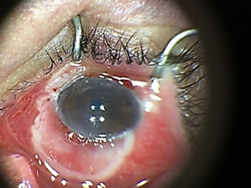
Mpox, caused by infection with Monkeypox virus, usually presents as a mild, self-limited illness in immunocompetent persons that resolves within 2-4 weeks. Serious complications have been reported when mpox lesions involve vulnerable anatomic sites, such as the eye, and in those with substantial immunosuppression. We describe a patient with advanced human immunodeficiency virus infection and sustained viral shedding of mpox with ocular involvement, which resulted in vision loss.
Keywords: HIV; MPOX; Ocular MPOX.
© The Author(s) 2023. Published by Oxford University Press on behalf of Infectious Diseases Society of America.
Conflict of interest statement
Potential conflicts of interest. All authors: No reported conflicts of interest.
Figures


References
-
- World Health Organization . Monkeypox.. Available at: https://www.who.int/health-topics/monkeypox/#tab=tab. Accessed 6 April 2023.
-
- Reed KD, Melski JW, Graham MB, et al. . The detection of monkeypox in humans in the Western Hemisphere. N Engl J Med 2004; 350:342–50. - PubMed
-
- European Centre for Disease Prevention and Control . Monkeypox multi-country outbreak situation update, 4 August 2022. Available at: ecdc.europa.eu/en/monkeypox-outbreak. Accessed 6 April 2023.
