

Subacute hemorrhagic pericardial tamponade after COVID-19 infection mimicking carcinomatous pericarditis: a case report
- PMID: 38264260
- PMCID: PMC10803410
- DOI: 10.3389/fcvm.2023.1329952
Subacute hemorrhagic pericardial tamponade after COVID-19 infection mimicking carcinomatous pericarditis: a case report
Abstract
Background: Coronavirus disease (COVID-19)-associated acute pericarditis has recently received much attention owing to its high frequency associated with pericardial tamponade (PT), showing unfavorable prognosis. However, early diagnosis and treatment remain challenging in cases of non-specific signs and symptoms.
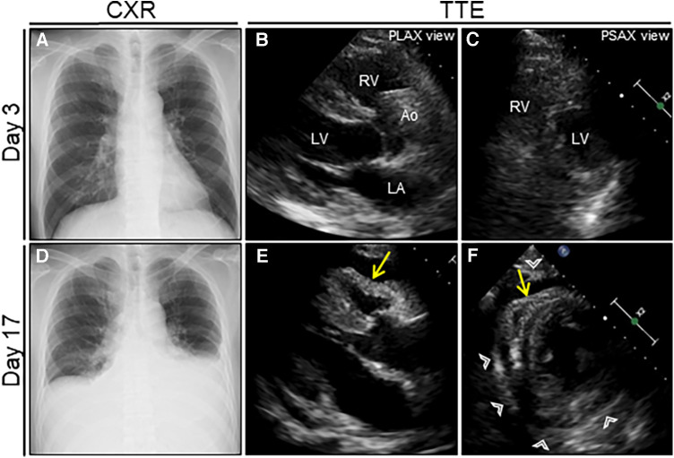
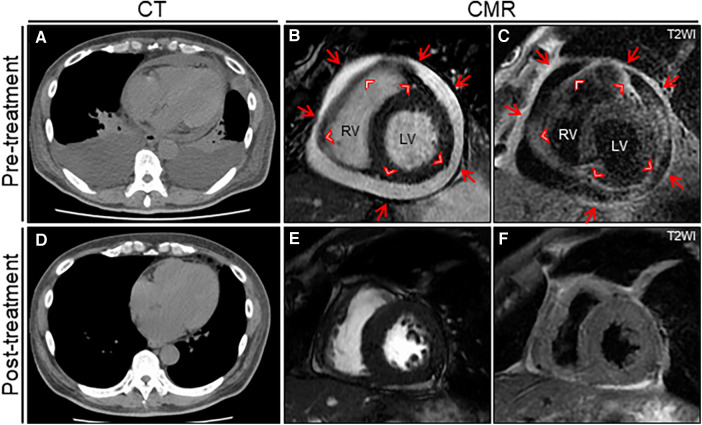
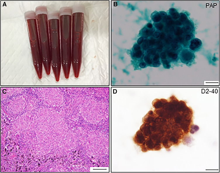
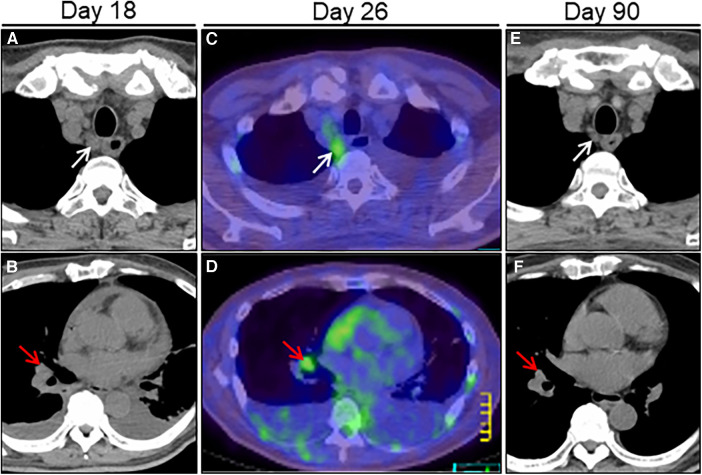
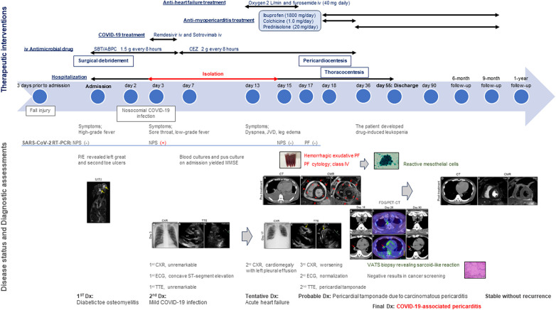
Case presentation: A 64-year-old man was admitted to our hospital for acute osteomyelitis of the toes and was properly treated with antimicrobial agents. Three days after admission, the patient developed mild COVID-19 without pneumonia, for which early anti-COVID-19 agents were initiated. Nevertheless, the patient developed hemorrhagic PT due to acute pericarditis 2 weeks later, which was confirmed by cardiac magnetic resonance, requiring an urgent pericardiocentesis. Although cytological analysis of the hemorrhagic pericardial fluid strongly suggested adenocarcinoma, the atypical cells were eventually proven to be mesothelial cells with reactive atypia. Furthermore, lymph nodes swelling with abnormal 2-[18F]-fluoro-2-deoxy-D-glucose accumulation on imaging were suggestive of malignancy. However, biopsy examination revealed multiple non-caseating granulomas in the lymph node, unlikely due to malignancy. Eventually, the temporal association of the preceding COVID-19 with the occurrence of subacute PT without other identifiable cause led to a final diagnosis of COVID-19-associated acute pericarditis. With anti-inflammatory and corticosteroids treatment, the patient's symptoms involving the pericardial structure and function were completely resolved along with improvements in size of the affected lymphadenopathies.
Conclusions: We encountered a unique case of COVID-19-associated acute pericarditis exhibiting hemorrhagic PT. This case underscores the residual risk of delayed pericardial involvement even in patients with mild COVID-19 who receive early treatment, and the recognition that COVID-19 may cause various cytomorphological and histological features. Additionally, the importance of considering this rare entity as a cause of hemorrhagic pericardial effusions should be highlighted.
Keywords: COVID-19; acute pericarditis; cytology; hemorrhagic pericardial tamponade; sarcoid-like reaction.
© 2024 Yamamoto, Kume, Hashimoto, Isogai, Kuwabara, Noguchi, Murayama, Hashimoto and Ogino.
Conflict of interest statement
The authors declare that the research was conducted in the absence of any commercial or financial relationships that could be construed as a potential conflict of interest.
Figures
Similar articles
-
Hemorrhagic Pericardial Effusion Secondary to Coxsackie B Pericarditis.Cureus. 2025 Jan 3;17(1):e76861. doi: 10.7759/cureus.76861. eCollection 2025 Jan. Cureus. 2025. PMID: 39897235 Free PMC article.
-
Diagnosis of malignant pericarditis: a single centre experience.Kardiol Pol. 2012;70(11):1147-53. Kardiol Pol. 2012. PMID: 23180523
-
Surviving the Storm: Cardiac Tamponade and Effusive Constrictive Pericarditis Complicated by Pericardial Decompression Syndrome Induced by COVID-19 Infection in the Setting of Newly Diagnosed Acute Myeloid Leukemia (AML).Cureus. 2024 Mar 22;16(3):e56710. doi: 10.7759/cureus.56710. eCollection 2024 Mar. Cureus. 2024. PMID: 38646402 Free PMC article.
-
[Toxoplasma pericarditis without immunosuppressant disorder detected by polymerase chain reaction of pericardial fluid: a case report].J Cardiol. 2000 Jan;35(1):47-54. J Cardiol. 2000. PMID: 10654250 Review. Japanese.
-
Management strategies in pericardial emergencies.Herz. 2006 Dec;31(9):891-900. doi: 10.1007/s00059-006-2937-0. Herz. 2006. PMID: 17180653 Review.
Cited by
-
From thrombosis to tamponade: unveiling severe pericardial effusion in a misdiagnosis case.Int J Emerg Med. 2025 Jan 3;18(1):4. doi: 10.1186/s12245-024-00794-z. Int J Emerg Med. 2025. PMID: 39754100 Free PMC article.
References
-
- Adler Y, Charron P, Imazio M, Badano L, Barón-Esquivias G, Bogaert J, et al. 2015 ESC guidelines for the diagnosis and management of pericardial diseases: the task force for the diagnosis and management of pericardial diseases of the European society of cardiology (ESC)Endorsed by: the European association for cardio-thoracic surgery (EACTS). Eur Heart J. (2015) 36(42):2921–64. 10.1093/eurheartj/ehv318 - DOI - PMC - PubMed
Publication types
LinkOut - more resources
Full Text Sources