

Area of the Fetal Ascending and Descending Aorta by Spatiotemporal Image Correlation in the Rendering Mode: Reproducibility and Comparison with Pregestational Diabetic Mothers
- PMID: 38264598
- PMCID: PMC10802868
- DOI: 10.4103/jmu.jmu_102_22
Area of the Fetal Ascending and Descending Aorta by Spatiotemporal Image Correlation in the Rendering Mode: Reproducibility and Comparison with Pregestational Diabetic Mothers
Abstract
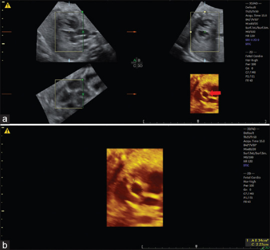
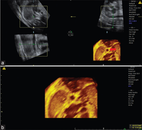
Background: The objective of this study was to assess the ascending and descending aorta area measurements by three-dimensional (3D) ultrasound using spatiotemporal image correlation (STIC) in the rendering mode comparing these measurements with pregestational diabetic mothers and assessing the reproducibility of the method.
Methods: We carried out a retrospective cross-sectional study with 58 normal and nine fetuses from pregestational diabetic mothers between 20 and 33 + 6 weeks of gestation. Fetal heart volumes were acquired at the level of four-chamber view to obtain the reconstructed planes for the ascending and descending aorta areas in the rendering mode. Linear regression was performed to assess the correlation between the fetal aorta areas and gestational age (GA). To assess the intra- and interobserver reproducibility, we used the concordance correlation coefficient (CCC).
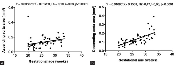
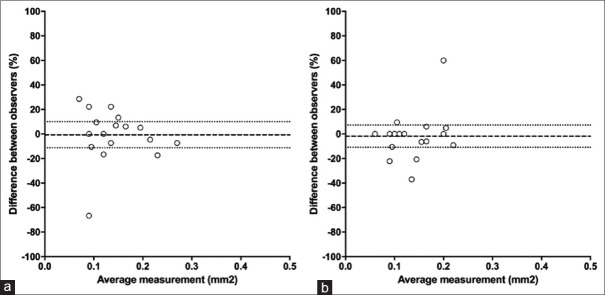
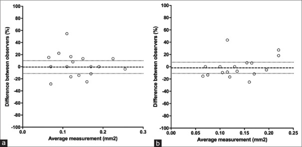
Results: The mean ascending and descending aorta areas were 0.12 (0.02-0.48) and 0.11 (0.04-0.39) cm2 in normal fetuses, respectively. There was a moderate positive correlation between GA and ascending aorta area measurements (0.005676*GA - 0.01283; r = 0.53, P < 0.0001) and strong positive correlation between GA and descending aorta area (0.01095*GA - 0.1581; r = 0.68, P < 0.0001). We observed a weak intra- and interobserver reproducibility with CCC ranging from 0.05 to 0.91. The mean difference in the ascending and descending aorta area measurements of normal and fetuses of pregestational diabetic mothers was -0.03 cm2 (P = 0.276) and -0.03 cm2 (P = 0.231), respectively.
Conclusion: The fetal ascending and descending aorta area measurements obtained by 3D ultrasound using STIC in the rendering mode increased with GA in normal fetuses. The method showed weak intra- and interobserver reproducibility.
Keywords: Aorta; area; fetal heart; pregestational diabetes; rendering mode; three-dimensional ultrasound.
Copyright: © 2023 Journal of Medical Ultrasound.
Conflict of interest statement
There are no conflicts of interest.
Figures

Similar articles
-
Fetal heart foramen ovale area by three-dimensional ultrasound using stic in the rendering mode: reference range and applicability in congenital heart diseases.Int J Cardiovasc Imaging. 2023 Mar;39(3):531-539. doi: 10.1007/s10554-022-02752-3. Epub 2022 Nov 5. Int J Cardiovasc Imaging. 2023. PMID: 36334212
-
References Values of Fetal Heart Myocardial Volume by Three-Dimensional Ultrasound using Spatiotemporal Image Correlation and Virtual Organ Computer-Aided Analysis Methods and Their Applicability in Pregestational Diabetic Women.Am J Perinatol. 2021 Jun;38(7):721-727. doi: 10.1055/s-0039-3400983. Epub 2019 Dec 13. Am J Perinatol. 2021. PMID: 31858500
-
Fetal tricuspid annular plane systolic excursion (f-TAPSE): evaluation of fetal right heart systolic function with conventional M-mode ultrasound and spatiotemporal image correlation (STIC) M-mode.Ultrasound Obstet Gynecol. 2013 Aug;42(2):182-8. doi: 10.1002/uog.12375. Ultrasound Obstet Gynecol. 2013. PMID: 23288668
-
Fetal cardiac function by mitral and tricuspid annular plane systolic excursion using spatio-temporal image correlation M-mode and left cardiac output in fetuses of pregestational diabetic mothers.Obstet Gynecol Sci. 2021 May;64(3):257-265. doi: 10.5468/ogs.20274. Epub 2021 Jan 27. Obstet Gynecol Sci. 2021. PMID: 33499582 Free PMC article.
-
Fetal Cardiac Function and Ventricular Volumes Determined by Three-Dimensional Ultrasound Using STIC and VOCAL Methods in Fetuses from Pre-gestational Diabetic Women.Pediatr Cardiol. 2020 Aug;41(6):1125-1134. doi: 10.1007/s00246-020-02362-7. Epub 2020 May 4. Pediatr Cardiol. 2020. PMID: 32367304
References
-
- Carlson BM. 6th ed. St. Louis, MO: Elservier; 2018. Human Embryology and Developmental Biology.
-
- Moore KL, Persaud TV, Torchia MG. 9th ed. Rio de Janeiro: Elservier; 2013. Clinical Embryology.
-
- Moore KL, Dalley AF, Agur AM. 8th ed. Philadelphia, PA: Wolters Kluwer; 2018. Clinically Oriented Anatomy.
-
- Evanoff NG, Dengel DR, Narasimhan S. Assessing vascular characteristics of the fetal descending aorta: A feasibility study. J Clin Ultrasound. 2020;48:211–5. - PubMed
LinkOut - more resources
Full Text Sources