

Case Reports
doi: 10.1016/j.case.2023.09.001.
eCollection 2024 Jan.
Double-Outlet Right Atrium in a Young Cat
Affiliations
- PMID: 38264619
- PMCID: PMC10801805
- DOI: 10.1016/j.case.2023.09.001
Item in Clipboard
Case Reports
Double-Outlet Right Atrium in a Young Cat
CASE (Phila).
.
No abstract available
Keywords: Endocardial cushion defect; Feline congenital cardiac defect; Feline echocardiography; Interatrial septum deviation.
Figures

Two-dimensional TTE, right parasternal long-axis 4-chamber view, demonstrates leftward deviation of the IAS (arrow) that appears as a shelf on the dorsal aspect of the mitral valve. The RA communicates with both ventricles, and there is a small ostium primum septal defect (∗) resulting in obstruction to the left atrial outflow.

Two-dimensional TTE, left apical 4-chamber view (aligned for assessment of LA outflow), pulsed-wave spectral Doppler, demonstrates increased blood flow velocity (∼1.7 m/sec) due to the leftward deviation of the IAS and the small ostium primum septal defect.

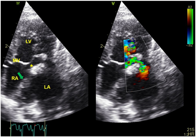
Two-dimensional TTE, left apical 4-chamber view without (left) and with (right) color-flow Doppler, demonstrates the flow obstruction to LA outflow caused by the leftward deviation of the IAS (arrow) and ostium primum defect (∗). MV, Mitral valve.

Dorsoventral (DV, A) and right lateral (RL, B) radiographic views of the thorax demonstrate a focal area of increased soft tissue opacity with an extrapleural sign between the cranial and caudal parts of the left cranial lung lobe (DV view) and a thin interlobar fissure between the cranial and caudal lung field (RL view). There is also a diffuse, mild interstitial lung pattern throughout the rest of the pulmonary parenchyma, and the pulmonary veins are dilated and taper peripherally. The cardiac silhouette is enlarged (vertebral heart size = 9 [reference interval, 6.7-8.1]).

Gross pathology image of a sagittal plane of the heart with a metallic probe (red arrow) passing from the LA into the LV through a small ostium primum defect and part of the extended RA. The IAS (white arrow) deviates leftward and dorsal to the mitral valve. AVC, AV canal.
Similar articles
-
Double-outlet right atrium in a 9 year-old cat.J Vet Cardiol. 2014 Jun;16(2):127-31. doi: 10.1016/j.jvc.2013.12.005. Epub 2014 Mar 1. J Vet Cardiol. 2014. PMID: 24747061
-
Double outlet right atrium in a dog.J Vet Cardiol. 2024 Aug;54:24-29. doi: 10.1016/j.jvc.2024.05.001. Epub 2024 May 16. J Vet Cardiol. 2024. PMID: 38851121
-
Diagnosis of endocardial cushion defect with cross-sectional and M-mode scanning echocardiography. Differentiation from secundum atrial septal defect.Br Heart J. 1976 Sep;38(9):911-20. doi: 10.1136/hrt.38.9.911. Br Heart J. 1976. PMID: 971376 Free PMC article.
-
Double-Outlet Right Atrium: Review of a Rare Anomaly With an Exemplary Case.World J Pediatr Congenit Heart Surg. 2020 Jan;11(1):79-84. doi: 10.1177/2150135119885895. World J Pediatr Congenit Heart Surg. 2020. PMID: 31835981 Review.
-
Essential Modifiers of Double Outlet Right Ventricle: Revisit With Endocardial Surface Images and 3-Dimensional Print Models.Circ Cardiovasc Imaging. 2018 Mar;11(3):e006891. doi: 10.1161/CIRCIMAGING.117.006891. Circ Cardiovasc Imaging. 2018. PMID: 29855425 Review.
References
-
- Lister A.L., Buchanan J.W. Radiographic and echocardio- graphic measurement of the heart in obese cats. Vet Radiol Ultrasound. 2000;41:320–325. - PubMed
-
- Scansen B.A., Schneider M., Bonagura J.D. Sequential segmental classification of feline congenital heart disease. J Vet Cardiol. 2015;17(Suppl 1):10–52. - PubMed
-
- Schrope D.P. Atrioventricular septal defects: natural history, echocardiographic, electrocardiographic, and radio- graphic findings in 26 cats. J Vet Cardiol. 2013;15:233–242. - PubMed
-
- Durham J., Maisenbacher H. Double-outlet right atrium in a 9 year-old cat. J Vet Cardiol. 2014;16:127–131. - PubMed
-
- Van Mierop L.H.S. In: Second Henry Ford Hospital International Symposium on Cardiac Surgery. Davila J.C., editor. Appleton-Century-Crofts; New York, NY: 1977. Pathology and pathogenesis of endocardial cushion defects. Surgical implications; pp. 201–207.
Publication types
LinkOut - more resources
Full Text Sources
Miscellaneous
