. 2024 Jan 15;137(2):jcs261408.
doi: 10.1242/jcs.261408. Epub 2024 Jan 31.

Role of BicDR in bristle shaft construction and support of BicD functions

Affiliations

Role of BicDR in bristle shaft construction and support of BicD functions

Aleksandra Jejina et al. J Cell Sci. .

Abstract

Cell polarization requires asymmetric localization of numerous mRNAs, proteins and organelles. The movement of cargo towards the minus end of microtubules mostly depends on cytoplasmic dynein motors. In the dynein-dynactin-Bicaudal-D transport machinery, Bicaudal-D (BicD) links the cargo to the motor. Here, we focus on the role of Drosophila BicD-related (BicDR, CG32137) in the development of the long bristles. Together with BicD, it contributes to the organization and stability of the actin cytoskeleton in the not-yet-chitinized bristle shaft. BicD and BicDR also support the stable expression and distribution of Rab6 and Spn-F in the bristle shaft, including the distal tip localization of Spn-F, pointing to the role of microtubule-dependent vesicle trafficking for bristle construction. BicDR supports the function of BicD, and we discuss the hypothesis whereby BicDR might transport cargo more locally, with BicD transporting cargo over long distances, such as to the distal tip. We also identified embryonic proteins that interact with BicDR and appear to be BicDR cargo. For one of them, EF1γ (also known as eEF1γ), we show that the encoding gene EF1γ interacts with BicD and BicDR in the construction of the bristles.

Keywords: Drosophila; BicD; BicD-related; Bicaudal-D; Bristle formation; Microtubule vesicle transport; Rab6; Spn-F.


Conflict of interest statement

Competing interests The authors declare no competing or financial interests.

Figures

Fig. 1.
Fig. 1.
Bristle phenotypes in BicDR mutants. (A) Macrochaetae of a wild-type control fly, the revertant w; BicDR(rev)/Df4515 (B) and w; BicDR71/Df4515 (C,D). Note that the notum of the w; BicDR(rev)/Df4515 control does not show any differences compared to the wild type, whereas w; BicDR71/Df4515 flies eclosed with shorter pSC macrochaetae (white arrows in D) and occasionally with an additional aSC bristle (blue arrow in D); identification of macrochaetae was according to Takano (1998). Arrow in C indicates a discolored bristle tip. Note that several of the mutant phenotype images are reproduced in Fig. 6 for ease of comparison. Images in A–D representative of 20–40 animals examined. (E) RNAi knockdown of BicDR induces defective bristles. w; UAS-BicDR-RNAi/en-Gal4; UAS-BicDR-RNAi/UAS-GFP. Note the slightly paler macrochaetae tips and occasional shorter macrochaetae (marked with an arrow). In A–E, thorax lengths were 1.0–1.1 mm. (F) More female flies displayed the bristle phenotype upon knockdown of BicDR (12 out of 18 female and 2 out of 8 male flies).
Fig. 2.
Fig. 2.
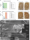
Effect of BicD and BicDR mutations on the structure of the macrochaetae. (A) Frequency of eclosed BicD; BicDR double mutants with the genotype w; BicDPA66/−; BicDR*/−. Adult flies eclosed only in crosses containing BicDR29 or the control BicDR(rev) except for one escaper with the allele BicDR71. All of them developed a short-bristle phenotype. The genotype w; BicDPA66/−; Df4515/+ eclosed in all crosses and all flies with the named genotype developed the short-bristle phenotype as well. The calculated expected frequency is shown in green. (B) Comparison of the bristle phenotypes observed in controls [white (w)], BicDPA66/− and BicDPA66/−; BicDR71/+ as well as BicDPA66/−; BicDRDf/+. Note that the BicD bristle phenotype, which manifests itself in discolored and brittle bristle tips, is stronger upon the reduction of BicDR function. The phenotypes observed in BicDPA66/−; BicDR71/+ and BicDPA66/−; BicDRDf/+ show the same severity, indicating that the allele BicDR71 behaves like a BicDRnull mutant for this phenotype. Arrow indicates a discolored bristle tip. Thorax lengths were 1.0–1.1 mm. (C) The frequency of the short bristle phenotype in BicDPA66/−; BicDR*/+ animals. The BicDR-excisions that the animals carry and the deficiency Df737 (this deficiency is also referred to as BicDRDf), are indicated. (D–H) Scanning electron micrographs of the posterior scutellar bristles (pSC) of the wild type, BicD and BicDR mutants and mutant combinations, showing their effects on bristle length and structure. Scale bars: 30 μm. (D) Wild type is an OreR line outcrossed to a white line. (E) BicDR mutants show slightly shorter, thinner bristles that appear flattened. (F) With only one normal copy of BicD, BicDR bristles are again slightly shorter. (G) BicDPA66/− bristles are only slightly shorter than wild-type bristles, but the enhancement of the phenotype by inactivating one copy of BicDR – shown with two unrelated alleles – is very strong (H). Note that in this genotype, the bundle structure is only visible close to the base and gets lost 50 μm distal to the base. ‘-’ indicates that the BicD or BicDR gene on the indicated chromosome was deleted by a small deficiency (Df7068 for BicD and Df4514 for BicDR). Image in D is representative of eight wild-type bristles. Image in E is representative of 16 bristles of the genotype BicDR71 over deficiency or BicDR8.1. Image in F is representative of 16 bristles of the genotype BicDR71 over deficiency or BicDR8.1. Image in G is representative of ten bristles of the genotype BicDRPA66 over deficiency. Images in H are representative of 12 (left) and eight (right) bristles.
Fig. 3.
Fig. 3.
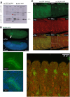
BicDR is expressed in the region of sensory organ precursors of stage 13 embryos. (A) Expression during the different stages of the life cycle is shown by a western blot stained for GFP to reveal the expression of the endogenously tagged BicDR (see Fig. S6 for uncropped images of the blot). The samples loaded were from stage 13 to 16 embryos, third-instar larvae, adult flies and salivary glands. All were of the genotype w; BicDR::GFP/Df4515 or the negative control (white). The loading control was GAPDH with a size of 35 kDa (see lower blot). BicDR-B::GFP, with a size of 130 kDa was found mostly in adult flies, 3rd instar larvae, and the dissected salivary glands of the third-instar larvae, while BicDR-A::GFP with 110 kDa was mostly expressed in late embryos. Blot is representative of two repeats. (B) Stage 13 embryos stained for BicDR::GFP (green). The DNA is stained with Hoechst (blue). BicDR::GFP is expressed apically in the cells of salivary glands and cells along the anterior-posterior embryo axis in a metameric pattern. Arrow highlights salivary gland. Magnified views are shown underneath. (C) Co-staining of BicDR::GFP embryos with the sensory organ precursor marker Asense (red) and GFP (green) identifies the GFP-positive cells in the vicinity of elevated Ase staining. Images in B and C are representative of five (B) and seven (C) repeats.
Fig. 4.
Fig. 4.
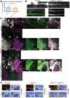
Pupal bristles depend on BicD and BicDR for actin bundle stability and proper localization of Spn-F and Rab6. (A) The length of single pupal macrochaetae was measured in white controls and BicDPA66/−; BicDR71/+ double mutants. No significant dissimilarities (ns) between the two groups were found at this stage of bristle development (two-tailed t-test). (B) Scutellar macrochaetae stained for F-actin and acetylated tubulin in controls and the indicated double mutant (gray, F-actin; green, acetylated tubulin). Images are representative of four (wild type) and seven (double mutant) repeats. (C) BicD and BicDR are needed to localize normal levels of Rab6 in the shaft of scutellar macrochaetae and at their bristle tips. This accumulation is impaired in the BicDPA66/− and particularly in the BicDPA66/−; BicDR71/+ double mutants (gray, F-actin; green, Spn-F; pink, Rab6; see also D and Figs S4, S5 for additional staining and relative quantification). The genotype of the sample is listed on the left side. All macrochaetae originate in the upper left corner (indicated with a ‘+’) and grow downwards to the lower right corner (indicated with a ‘–’) and are highlighted by orange dashed lines. The tip is visualized with staining for Spn-F. The enlargement of the bristle tips framed by the red boxes in C is shown for the three channels. The outlines of the bristle cells were estimated from the F-actin staining and the staining for Spn-F and Rab6. The localization of Rab6 decreases toward the bristle tip of BicD mutants, whereas it was completely absent in the distal tips of the BicD; BicDR double mutants. (D) Intensity plots of Rab6 and Spn-F signals in each image plane to visualize the distribution of Rab6 and Spn-F signals through the bristle shaft. The highest Z-score of 4 is shown in red; dark blue marks a Z-score of 0 and indicates that no signal could be detected. The segmented line drawn through the bristle shaft has a width of 10 pixels and their mean result was used for the graphs. Images in D are representative examples for five repeats each.
Fig. 5.
Fig. 5.
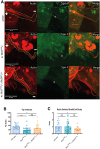
Pupal bristles of BicD; BicDR double mutants show impaired Spn-F localization at the bristle tip. (A) Stained macrochaetae (red, F-actin; green, Spn-F). The genotypes of the samples are listed on the left side. All macrochaetae originate in the upper left corner and have their tips pointing downwards (bristles are outlined by the dashed yellow line, + is the proximal end, − the distal end). The localization of Spn-F at the bristle tip is much weaker in w; BicDPA66/Df7068; TM6B/+ and w; BicDPA66/Df7068; BicDR71/+ pupae. This observation could be confirmed with the calculation of the tip index shown in B. (B) The average tip index of the mutants is significantly lower than those of the control group. (C) The ratio of Spn-F signal in the elongated bristle shaft versus its cell body is significantly lower in BicDPA66/−; BicDR*/+ animals. White squares indicate the positions within the bristle shaft where the signal intensity of Spn-F was measured. For the calculation, the signal was measured within one plane in the approximately middle part of the bristle shaft and divided through the signal intensity measured within the plane where the actin bundles sprout out of the tissue. Error bars are s.d. In B and C, n=19 (wild type), 23 (BicDPA66/–) and 24 (BicDPA66/–; BicDR71/+). *P<0.05; ***P<0.001; ns, not significant (one-way ANOVA with Dunnett's multiple comparisons test).
Fig. 6.
Fig. 6.
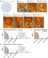
Genetic interaction between BicDR and EF1γ in bristle construction. (A) Proteins identified in the cut-out gel bands from the tagged BicDR and BicDRK555A immunoprecipitations. A total of 285 potential binding partners were identified; 82 proteins were found in both samples, whereas 179 proteins were found only in the wild-type BicDR::GFP IP. (B–H) Resemblance of phenotypes compared to the (B) white control (0% short bristle phenotypes; n=21), (C) BicDR(rev)/− control (0% short bristle phenotype; n=13), (D) BicDR71/− (35% of flies displayed such a bristle phenotype; n=11), (E) EF1γA70/− (35% of flies displayed a short bristle phenotype; n=9), (F) EF1γA42/− (46% of flies eclosed with additional bristles; n=11), (G) EF1γA28/− (50% of flies had additional and shorter bristles; n=26), (H) EF1γA15/− (50% of flies had shorter bristles; n=18). Note that the white control and 93% of the BicDR revertants, BicDR(rev)/−, eclosed without a visible bristle phenotype. A total of 7% of BicDR(rev)/− animals contained an additional aSC bristle. Flies with the genotypes BicDR71/−, EF1γA70/−, and EF1γA51/− showed shorter pSC macrochaetae. Additionally, 21% of BicDR71/− animals eclosed with an extra aSC bristle, a similar frequency to that observed with the alleles EF1γA42/− and EF1γA28/−. Arrows point to shorter posterior macrochaetae (D,E,G and H) and to more anterior bristle duplications (D,F and G). Note that several of the mutant phenotype images shown in Fig. 1 are reproduced here for ease of comparison. Thorax lengths were 1.0–1.1 mm. Images are representative of three repeats. (I–K) Effect of combining heterozygous EF1γA70, EF1γA42 or EF1γA28 with heterozygous BicD and BicDR alleles showing strong genetic interactions between heterozygous BicD, BicDR, and EF1y alleles. Frequency of mutant phenotypes observed in double and triple heterozygous combinations. Different mutant combinations containing a BicDR*, BicDPA66, and EF1γ* allele eclosed with different bristle phenotypes. The frequency of animals that eclosed with a short-bristle phenotype is significantly higher if the animals carry a BicDPA66 and BicDR71 allele except for the combination with EF1γA70 where the frequency of flies with short bristles was the highest in BicDPA66/+; EF1γA70/+ (30%). Noticeable is that 19% of the flies with the genotype BicDPA66/+; BicDR71/EF1γA28 eclosed with short bristles, even though the allele EF1γA28 /− induces additional bristles. n=44–84 (I), 37–95 (J) and 52–78 (K).

Update of

Similar articles

References

    1. Abdelilah-Seyfried, S., Chan, Y. M., Zeng, C., Justice, N. J., Younger-Shepherd, S., Sharp, L. E., Barbel, S., Meadows, S. A., Jan, L. Y. and Jan, Y. N. (2000). A gain-of-function screen for genes that affect the development of the drosophila adult external sensory organ. Genetics 155, 733-752. 10.1093/genetics/155.2.733 - DOI - PMC - PubMed
    1. Abdu, U., Bar, D. and Schüpbach, T. (2006). spn-F encodes a novel protein that affects oocyte patterning and bristle morphology in Drosophila. Development 133, 1477-1484. 10.1242/dev.02319 - DOI - PubMed
    1. Adler, P. N. (2020). The localization of chitin synthase mediates the patterned deposition of chitin in developing Drosophila bristles. bioRxiv 718841. 10.1101/718841 - DOI
    1. Aradska, J., Bulat, T., Sialana, F. J., Birner-Gruenberger, R., Erich, B. and Lubec, G. (2015). Gel-free mass spectrometry analysis of Drosophila melanogaster heads. Proteomics 15, 3356-3360. 10.1002/pmic.201500092 - DOI - PubMed
    1. Bagley, J. A., Yan, Z., Zhang, W., Wildonger, J., Jan, L. Y. and Jan, Y. N. (2014). Double-Bromo and Extraterminal (BET) domain proteins regulate dendrite morphology and mechanosensory function. Genes Dev. 28, 1940-1956. 10.1101/gad.239962.114 - DOI - PMC - PubMed

Publication types

MeSH terms