. 2024 Jan 24;19(1):e0297516.
doi: 10.1371/journal.pone.0297516. eCollection 2024.

The spike protein of the apathogenic Beaudette strain of avian coronavirus can elicit a protective immune response against a virulent M41 challenge

Affiliations

The spike protein of the apathogenic Beaudette strain of avian coronavirus can elicit a protective immune response against a virulent M41 challenge

Sarah Keep et al. PLoS One. .

Abstract

The avian Gammacoronavirus infectious bronchitis virus (IBV) causes major economic losses in the poultry industry as the aetiological agent of infectious bronchitis, a highly contagious respiratory disease in chickens. IBV causes major economic losses to poultry industries across the globe and is a concern for global food security. IBV vaccines are currently produced by serial passage, typically 80 to 100 times in chicken embryonated eggs (CEE) to achieve attenuation by unknown molecular mechanisms. Vaccines produced in this manner present a risk of reversion as often few consensus level changes are acquired. The process of serial passage is cumbersome, time consuming, solely dependent on the supply of CEE and does not allow for rapid vaccine development in response to newly emerging IBV strains. Both alternative rational attenuation and cell culture-based propagation methods would therefore be highly beneficial. The majority of IBV strains are however unable to be propagated in cell culture proving a significant barrier to the development of cell-based vaccines. In this study we demonstrate the incorporation of a heterologous Spike (S) gene derived from the apathogenic Beaudette strain of IBV into a pathogenic M41 genomic backbone generated a recombinant IBV denoted M41K-Beau(S) that exhibits Beaudette's unique ability to replicate in Vero cells, a cell line licenced for vaccine production. The rIBV M41K-Beau(S) additionally exhibited an attenuated in vivo phenotype which was not the consequence of the presence of a large heterologous gene demonstrating that the Beaudette S not only offers a method for virus propagation in cell culture but also a mechanism for rational attenuation. Although historical research suggested that Beaudette, and by extension the Beaudette S protein was poorly immunogenic, vaccination of chickens with M41K-Beau(S) induced a complete cross protective immune response in terms of clinical disease and tracheal ciliary activity against challenge with a virulent IBV, M41-CK, belonging to the same serogroup as Beaudette. This implies that the amino acid sequence differences between the Beaudette and M41 S proteins have not distorted important protective epitopes. The Beaudette S protein therefore offers a significant avenue for vaccine development, with the advantage of a propagation platform less reliant on CEE.


Conflict of interest statement

I have read the journal’s policy and the authors of this manuscript have the following competing interests: A patent has previously been filed by The Pirbright Institute to protect the intellectual property of the work surrounding mutations Pro85Leu in Nsp 10 and Val393Leu in Nsp 14 (Patent name: Coronavirus, Number EP3172319B1, Authors: Erica Bickerton, Sarah Keep and Paul Britton). The authors declare no additional conflicts of interest. This does not alter our adherence to PLOS ONE policies on sharing data and materials.

Figures

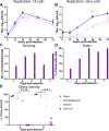
Fig 1
Fig 1. The incorporation of a heterologous S gene has resulted in altered tropism in vitro.
(A, B, E, F) Ex vivo TOCs, (C) CK cells and (D) Vero cells were inoculated with 104 PFU or CD50 equivalent dose of either (A-D) M41-K, Beau-R or M41K-Beau(S) or (E-F) of M41-K, 4/91 or M41K-4/91(S). (A, C, D, E) Supernatant was collected at 24-hour intervals and titrated on (A, C, D) CK cells or (E) in ex vivo TOCs. (B, F) The percentage ciliary activity was assessed at 24-hour intervals. Note: TOCs were inoculated in replicates of 10, and mock infected TOCs were inoculated with media only. (A–F) Each data point represents the average titre from three independent experiments with error bars representing SEM. Statistical differences, * p<0.05 and ** p<0.05 were assessed by Two-Way ANOVA followed by Tukey’s test for multiple comparisons. (B, F) Statistical differences identified were between all viruses and mock from 24 to 96 hpi; no differences were identified between the viruses. (A, C, D) Only statistical differences between M41K-Beau(S) and M41-K are displayed. Additional statistical differences are as follows: (C) M41-K vs Beau-R, 24 hpi and (D) M41-K vs Beau-R, 24–96 hpi.
Fig 2
Fig 2. M41K-Beau(S) is attenuated in vivo.
At 8 days of age, four groups of (A-C) 12 or (D-F) 15 SPF RIR chicks were inoculated via the intraocular and intranasal route with 105 PFU or equivalent CD50 dose of either (A-C) M41K-Beau(S), M41-K or M41-CK or (D-F) with M41K-4/91(S), M41-K or 4/91. Mock infected birds were inoculated with PBS. Clinical signs were observed from either day 2 or 3 to day 7 post-infection. (A, D) The average number of snicks per bird per minute was calculated for each group based on counts from two persons. (B, E) The percentage of birds exhibiting rales in each group was calculated. (C, F) Ciliary activity in the trachea was measured on days 4 and 6 post-infection. Trachea were extracted from three or five randomly selected birds on each day and sectioned into 10 x 1 mm rings. Each ring was analysed under a light microscope and assigned a score between 1 and 4 depending on the proportion of cilia beating. 1 = 25% beating, 2 = 50%, 3 = 75%, 4 = 100%. Each data point represents the average percentage ciliary activity of each bird. Error bars represent SD. Statistical differences were assessed using a Two-Way ANOVA followed by Tukey’s test for multiple comparisons and are highlighted by ***(p<0.0005) and **** (p<0.0001).
Fig 3
Fig 3. Viral load in trachea and eyelid tissue is comparable between M41K-Beau(S) and M41-K.
Samples of homogenised (A–C) eyelid and (D–F) trachea harvested on days 4 and 6 post-infection for experiment 1 and day 4 only for experiment 2 were titrated in ex vivo TOCs. Unfortunately, no tissue was available from day 6 in experiment 2 for this assay. Viral load in terms of CD50 was calculated for each sample using the Reed-Muench method. Statistical differences were identified using a One-Way ANOVA followed by Tukey’s test for multiple comparisons and are highlighted by * (p<0.05) and ** (p<0.005). Of note, one sample of eyelid was damaged during extraction and there was not enough tissue available for this assay therefore in panel C there are only 4 birds for M41K-4/91(S).
Fig 4
Fig 4. M41R-Beau(S) is attenuated in vivo.
CK cells (A) and Vero cells (B) were inoculated with either 0.5 x 10³ PFU of M41R-Beau(S), Beau-R or M41-R (MOI ~ 0.001). Supernatant was harvested and titrated on CK cells. Error bars represent SEM of three independent repeats. Statistical differences were analysed using a Two-way ANOVA with a Tukey’s test for multiple comparisons. Differences identified are (A) Beau-R vs M41-R 72 hpi; (B) Beau-R vs M41-R, 24–96 hpi and M41R-Beau(S) vs M41-R and vs Beau-R at 48 hpi (*p<0.05, **p<0.005). (C- E) Groups of 12 SPF RIR chickens at 8 days of age were inoculated with 105 PFU of M41R-Beau(S), M41-R or M41-CK or mock inoculated with PBS via the intraocular and intranasal route. Clinical signs were observed from day 3 to 7 post-infection. (C) The average number of snicks per bird per minute was calculated for each group based on counts from two persons. (D) The percentage of birds exhibiting rales in each group was calculated. (E) Trachea were extracted from three randomly selected birds on each day and sectioned into 10 x 1 mm rings. Each ring was analysed under a light microscope and assigned a score between 1 and 4 depending on the proportion of cilia beating. 1 = 25% beating, 2 = 50%, 3 = 75%, 4 = 100%. Each data point represents the average percentage ciliary activity of each bird. Error bars represent SD. Statistical differences were assessed using a Two-Way ANOVA followed by Tukey’s test for multiple comparisons and are highlighted by **** (p<0.0001).
Fig 5
Fig 5. Vaccination with M41K-Beau(S) or M41R-Beau(S) protects against M41-CK challenge.
Groups of 30 SPF RIR chicks were vaccinated at 8 days old with 104 PFU of either M41K-Beau(S) or M41R-Beau(S) or mock vaccinated with PBS via the intraocular and intranasal route. At 27 days post-vaccination chickens were either challenged with 104 M41-CK or mock challenged with PBS also via the intranasal and intraocular route. Clinical signs including snicking (A and B) and rales (C and D) were observed from day 3 to day 7 post-vaccination and day 3 to 7 post-challenge. Trachea were extracted from six randomly selected birds on day 4 post-vaccination (E), day 4 and 6 post-challenge (F) and sectioned into 10 x 1 mm rings. Each ring was analysed under a light microscope and assigned a score between 1 and 4 depending on the proportion of cilia beating. 1 = 25% beating, 2 = 50%, 3 = 75%, 4 = 100%. Each data point represents the average percentage ciliary activity of each bird. Error bars represent SD. Statistical differences were assessed using a Two-Way ANOVA followed by Tukey’s test for multiple comparisons and are highlighted by **** (p<0.0001).
Fig 6
Fig 6. Vaccination with M41K-Beau(S) and M41R-Beau(S) induces a robust antibody response.
Serum was harvested from chickens on day 14 post-vaccination (A) and days 4 (B) and 14 post-challenge (C) and diluted 1/80 for ELISA using the commercial BioChek IBV ELISA kit. Samples from each bird in a group harvested on day 14 post-vaccinated were pooled together due to the limited quantities of serum able to be collected. Samples were run in triplicate, and the average S/P ratio was calculated for each bird. S/P ratios for each independent repeat (A) or for each bird (B and C) are displayed with error bars representing the SD. (D) To assess the levels of neutralizing antibody, serum from day 14 post-challenge was serially diluted and incubated with 103 PFU of M41-CK, followed by a plaque assay on CK cells to determine the plaque reduction neutralization titer (PRNT50). The PRNT50 values were calculated for each group using the Reed-Muench method. Average PRNT50 values for each bird are displayed, with error bars representing the SD. Statistical differences were assessed by one-way ANOVA, followed by Tukey’s test for multiple comparisons and are highlighted by * (p<0.05), *** (p< 0.0005) and **** (p<0.0001).

References

    1. Cook JKA, Jackwood M, Jones RC. The long view: 40 years of infectious bronchitis research. Avian Pathol. 2012;41(3):239–50. doi: 10.1080/03079457.2012.680432 - DOI - PubMed
    1. Boursnell MEG, Brown TDK, Foulds IJ, Green PF, Tomley FM, Binns MM. Completion of the sequence of the genome of the coronavirus avian infectious bronchitis virus. Journal of General Virology. 1987;68(1):57–77. doi: 10.1099/0022-1317-68-1-57 - DOI - PubMed
    1. da Silva AP, Hauck R, Nociti SRC, Kern C, Shivaprasad HL, Zhou H, et al.. Molecular Biology and Pathological Process of an Infectious Bronchitis Virus with Enteric Tropism in Commercial Broilers. Viruses. 2021;13(8). doi: 10.3390/v13081477 - DOI - PMC - PubMed
    1. Najimudeen SM, Abd-Elsalam RM, Ranaweera HA, Isham IM, Hassan MSH, Farooq M, et al.. Replication of infectious bronchitis virus (IBV) Delmarva (DMV)/1639 variant in primary and secondary lymphoid organs leads to immunosuppression in chickens. Virology. 2023;587:109852. doi: 10.1016/j.virol.2023.109852 - DOI - PubMed
    1. Isham IM, Hassan MSH, Abd-Elsalam RM, Ranaweera HA, Mahmoud ME, Najimudeen SM, et al.. Impact of Maternal Antibodies on Infectious Bronchitis Virus (IBV) Infection in Primary and Secondary Lymphoid Organs of Chickens. Vaccines (Basel). 2023;11(7). doi: 10.3390/vaccines11071216 - DOI - PMC - PubMed