. 2024 Jan 30;121(5):e2304680121.
doi: 10.1073/pnas.2304680121. Epub 2024 Jan 24.

Expression of Atoh1, Gfi1, and Pou4f3 in the mature cochlea reprograms nonsensory cells into hair cells

Affiliations

Expression of Atoh1, Gfi1, and Pou4f3 in the mature cochlea reprograms nonsensory cells into hair cells

Melissa M McGovern et al. Proc Natl Acad Sci U S A. .

Abstract

Mechanosensory hair cells of the mature mammalian organ of Corti do not regenerate; consequently, loss of hair cells leads to permanent hearing loss. Although nonmammalian vertebrates can regenerate hair cells from neighboring supporting cells, many humans with severe hearing loss lack both hair cells and supporting cells, with the organ of Corti being replaced by a flat epithelium of nonsensory cells. To determine whether the mature cochlea can produce hair cells in vivo, we reprogrammed nonsensory cells adjacent to the organ of Corti with three hair cell transcription factors: Gfi1, Atoh1, and Pou4f3. We generated numerous hair cell-like cells in nonsensory regions of the cochlea and new hair cells continued to be added over a period of 9 wk. Significantly, cells adjacent to reprogrammed hair cells expressed markers of supporting cells, suggesting that transcription factor reprogramming of nonsensory cochlear cells in adult animals can generate mosaics of sensory cells like those seen in the organ of Corti. Generating such sensory mosaics by reprogramming may represent a potential strategy for hearing restoration in humans.

Keywords: cochlea; hair cells; inner ear; reprogramming.


Conflict of interest statement

Competing interests statement:The authors declare no competing interest.

Figures

Fig. 1.
Fig. 1.
Nonsensory cells of the mature cochlea are reprogrammed into hair cell–like cells by expression of Atoh1, Gfi1, and Pou4f3. (A) Timeline for the activation of reprogramming factors and reporter expression. TMX was injected on two consecutive days at 3 wk of age. Cochleas were collected and analyzed at 5 wk of age. (B) The reprogramming factors and/or tdTomato reporter were expressed in nonsensory cells of the IS and OS. Hair cells are detected by Myosin VIIa staining (green) in the Fbxo2CreERT2::Rosa26tdTomato control cochlea. Hair cells are observed only in the organ of Corti, while tdTomato (red) was detected medial and lateral to the organ of Corti (n = 4). (C) Atoh1 activation alone (Fbxo2CreERT2::RosatdTomato::RosaA) is insufficient to reprogram nonsensory cells into hair cell–like cells (n = 8); however (D and D’) following expression of Atoh1+Gfi1+Pou4f3 (Fbxo2CreERT2:: RosaotdTomato:: RosaGAP), hair cells (green) are observed in the SL and OS (n = 19). (E) Quantification of ectopic hair cells in the IS and SL of Fbxo2CreERT2:: RosatdTomato:: RosaA (Atoh1) and Fbxo2CreERT2:: RosatdTomato:: RosaGAP (Gfi1+Atoh1+Pou4f3) cochleas (n = 3). (F) The hair cell protein, Parvalbumin (magenta), is limited to endogenous hair cells (green) in the organ of Corti in the control cochlea (Fbxo2CreERT2:: RosatdTomato, n = 4) but is expressed in reprogrammed hair cells in the SL following Atoh1+Gfi1+Pou4f3 expression (Fbxo2CreERT2:: RosatdTomato:: RosaGAP, n = 4). (G) Hair cells (green) were limited to the organ of Corti in the control cochlea in all cochlear turns. Following reprogramming with Atoh1, Gfi1, and Pou4f3, Myosin VIIa–positive cells were observed in nonsensory regions throughout the cochlear length. More hair cells were reprogrammed in the apex compared to the middle or basal turns of the cochlea. n = 3, *P < 0.05, **P < 0.01; (scale bar in AD’, F, and G = 50 µm), OS = outer sulcus, OoC = organ of Corti, IS = inner sulcus, SL = spiral limbus.
Fig. 2.
Fig. 2.
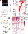
Single-cell RNA sequencing reveals that nonsensory cells activate hair cell networks following expression of Atoh1, Gfi1, and Pou4f3. (A) Representative image illustrating the expression pattern of Rosa26tdTomato (Red) when induced by Fbxo2CreERT2 and the epithelial marker EpCAM (Blue) at 5 wk. OS = outer sulcus, OoC = organ of Corti, IS = inner sulcus, SL = Spiral limbus. Fbxo2CreERT2::Rosa26-tdTomato (control) and Fbxo2CreERT2::Rosa26tdTomato::Rosa26-GAP cochleas were dissociated into single cells, and (B) tdTomato+EpCAM double positive cells were sorted by flow cytometry and collected for single-cell RNA sequencing. (C) Unbiased Seurat analysis revealed that control cells (blue) and reprogrammed cells (gold) largely clustered based on condition and 9 unique clusters were identified. RNA-velocity predicts that cells are transitioning from control cells into reprogrammed cells. (D) Known hair cell genes were activated at a higher level in one cluster of reprogrammed cells (Rprg HCs) than in two other reprogrammed clusters (Rprg Orig SCs and Rprg New SCs). (E) The heatmap showing the top 100 hair cell–specific genes, found previously at P1 from ref. , in reprogrammed cells. (F) SCENIC analysis detected the activation of genes associated with Atoh1 and Pou4f3 regulons in reprogrammed hair cells (Rprg HCs). (Scale bar in A, 50 µm.)
Fig. 3.
Fig. 3.
Reprogrammed hair cells signal to their neighbors to activate supporting cell genes. (A) Supporting cell genes are expressed in original supporting cells (Ctrl SCs) and are maintained in reprogrammed original supporting cells (Rprg Orig SCs). These genes are also activated in nonsensory cells following reprogramming (Rprg New SCs). (B) Sox2 (magenta) is restricted to supporting cells of the control cochlea (n = 4). Following reprogramming, Sox2 is expressed in cells of the SL, and some Sox2+/Myosin VIIa–cells were detected (arrowheads, n = 9). More Sox2-positive/Myosin VIIa–negative cells were observed in the apex compared to the middle and basal turns. *P < 0.05. OS = outer sulcus, OoC = organ of Corti, IS = inner sulcus, SL = spiral limbus. (C) The proliferation marker Ki-67 was not observed in the control cochlea; however, a small number of Ki-67-positive cells were observed following reprogramming. (D) The Notch receptor Notch1 is expressed in both control and reprogrammed cells. The Notch ligands Jagged 2 (Jag2), Delta like ligand 3 (Dll3), and Delta like ligand 2 (Dll2) are expressed in some reprogrammed cells. The Notch effector Hes5, which is associated with the promotion of supporting cell identity, is also expressed in some reprogrammed cells. (Scale bar in B, 50 µm and 20 µm.)
Fig. 4.
Fig. 4.
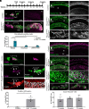
Hair cell–like cells are added over the course of 9 wk following the expression of Atoh1, Gfi1, and Pou4f3. (A) Reprogramming was activated at 3 wk of age in nonsensory cells of the inner ear and cochlea were analyzed at 5, 8, or 12 wk of age. (B) In the control cochlea, the normal complement of inner and outer hair cells (green) was observed n = 4. At 5 wk, Myosin VIIa+ hair cells were observed in the SL and OS in the apical turn and the density of these cells declined toward the basal turn. At 8 wk, the density of hair cells had increased in the SL and OS throughout the length of the cochlea. At 12 wk, cells throughout the length of the cochlea, in the SL, the IS, and the OS are robustly converted into hair cell–like cells. (C) Quantification of hair cells per 200 µm region of the IS and SL of Atoh1 expressing cochlea at 5 and 8 wk and Atoh1+Gfi1+Pou4f3 expressing cochlea at 5, 8, and 12 wk of age. (D) Quantification cells in the apical turn comparing the medial compartment and lateral compartment of control or Atoh1+Gfi1+Pou4f3 expressing cochlea at 12 wk of age. (E) Higher magnification images of Atoh1+Gfi1+Pou4f3 expressing cochlea at 8 and 12 wk of age. Quantification of reprogrammed hair cells per 200 µm reveals that there is an increase in the number of reprogrammed cells between 5 and 8 wk of age in the middle and base, while there is an increase in the apex between 8 and 12 wk of age. *P < 0.05, **P < 0.0, ***P < 0.001, and ****P < 0.0001.
Fig. 5.
Fig. 5.
Reprogrammed cells express multiple mature hair cell genes. (A) Reprogramming was activated at 3 wk of age in nonsensory cells of the inner ear and cochlea were analyzed at 5, 8, or 12 wk of age. (B) Parvalbumin (magenta) continues to be expressed in Rprg HCs in the SL and inner sulcus. Quantification of Parvalbumin-positive cells reveals that there is a significant increase in positive cells in the medial compartment (medial to the tunnel of Corti) but not in the Organ of Corti or floor of the cochlear duct or lateral wall (n = 3). (C) Prestin (magenta) is expressed in outer hair cells in the control cochlea and is expressed in some Rprg HCs (determined by the colabeling of Myosin VIIa in green and tdTomato in red) adjacent to the organ of Corti. The organ of Corti is disorganized following reprogramming, and the arrow indicates an endogenous hair cell and arrowheads indicate Rprg HCs. Significantly more Prestin-positive/tdTomato-positive cells were observed following reprogramming (n = 3). (D) Phalloidin labeling (white) is limited to the organ of Corti in the control cochlea (n = 6); however, numerous phalloidin+ cells are observed in the SL, IS, and OS following reprogramming (n = 2). Higher magnification analysis reveals distinct stereocilia atop Rprg HCs. (E) Sox2 expression (magenta) is limited to the organ of Corti in the control cochlea but is detected in the SL, Is, and OS following reprogramming (n = 2). Several cells were observed to be Sox2-positive and Myosin VIIa–negative (green; arrowheads). There was not a significant increase in the number of Sox2-positive/Myosin VIIa–negative cells with increased duration of reprogramming. Inner hair cells are indicated by asterisks, and outer hair cells are indicated with brackets in A and CE. TMX = tamoxifen, OHCs = outer hair cells, IHCs = inner hair cells. (Scale bar in B, 200 µm; (C and D) low mag = 20 µm; E low mag = 50 µm; E med mag = 10 µm, high mag = 2 µm.) OS = outer sulcus, OoC = organ of Corti, IS = inner sulcus, SL = spiral limbus. *P < 0.05 and ***P < 0.001.

Similar articles

Cited by

References

    1. Le T. N., Straatman L. V., Lea J., Westerberg B., Current insights in noise-induced hearing loss: A literature review of the underlying mechanism, pathophysiology, asymmetry, and management options. J. Otolaryngol. Head Neck Surg. 46, 1–15 (2017). - PMC - PubMed
    1. Henley C. M., Rybak L. P., Ototoxicity in developing mammals. Brain Res. Rev. 20, 68–90 (1995). - PubMed
    1. Walters B. J., et al. , In vivo Interplay between p27Kip1, GATA3, ATOH1, and POU4F3 converts non-sensory cells to hair cells in adult mice. Cell Rep. 19, 307–320 (2017). - PMC - PubMed
    1. Kraft S., Hsu C., Brough D. E., Staecker H., Atoh1 induces auditory hair cell recovery in mice after ototoxic injury. Laryngoscope 123, 992–999 (2013). - PMC - PubMed
    1. Yang S. M., et al. , Regeneration of stereocilia of hair cells by forced Atoh1 expression in the adult mammalian cochlea. PLoS One 7, e46355 (2012). - PMC - PubMed

Substances