. 2024 Feb 16;13(2):466-473.
doi: 10.1021/acssynbio.3c00682. Epub 2024 Jan 24.

Engineering an Autonucleolytic Mammalian Suspension Host Cell Line to Reduce DNA Impurity Levels in Serum-Free Lentiviral Process Streams

Affiliations

Engineering an Autonucleolytic Mammalian Suspension Host Cell Line to Reduce DNA Impurity Levels in Serum-Free Lentiviral Process Streams

Geoffrey Howe et al. ACS Synth Biol. .

Abstract

We engineered HEK293T cells with a transgene encoding tetracycline-inducible expression of a Staphylococcus aureus nuclease incorporating a translocation signal. We adapted the unmodified and nuclease-engineered cell lines to grow in suspension in serum-free media, generating the HEK293TS and NuPro-2S cell lines, respectively. Transient transfection yielded 1.19 × 10⁶ lentiviral transducing units per milliliter (TU/mL) from NuPro-2S cells and 1.45 × 10⁶ TU/mL from HEK293TS cells. DNA ladder disappearance revealed medium-resident nuclease activity arising from NuPro-2S cells in a tetracycline-inducible manner. DNA impurity levels in lentiviral material arising from NuPro-2S and HEK293TS cells were undetectable by SYBR Safe agarose gel staining. Direct measurement by PicoGreen reagent revealed DNA to be present at 636 ng/mL in lentiviral material from HEK293TS cells, an impurity level reduced by 89% to 70 ng/mL in lentiviral material from NuPro-2S cells. This reduction was comparable to the 23 ng/mL achieved by treating HEK293TS-derived lentiviral material with 50 units/mL Benzonase.

Keywords: bioprocess; gene therapy; lentivirus; mammalian cells; nuclease.


Conflict of interest statement

The authors declare no competing financial interest.

Figures

Figure 1
Figure 1
Anticipated compatibility of nuclease secretion and lentivirus production. Transgenes encoding lentiviral genomes and proteins are distributed across four plasmids (yellow circles) used for transient transfection of host cells. Complete lentiviral particles then assemble in the cytosol and, at the cell membrane, bud from the cell, taking a section of the membrane. Nuclease expressed from a genomically integrated transgene possesses a translocation signal intended to ensure that the recombinant enzyme is sequestered within the lumen of the endoplasmic reticulum (ER) and then trafficked in vesicles through the secretory pathway into the external milieu. Thus, host cell nucleic acids and lentiviral genomes are never in the same subcellular compartment as nuclease activity. Created with BioRender.
Figure 2
Figure 2
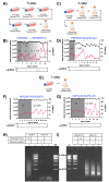
Adapting adherent cells to serum-free growth in suspension. (A) A five-step method in which v/v serum percentage was reduced stepwise in static culture before transfer to serum-free media in shake flasks. (B) Plots showing increase in viability metrics and suspension-mode growth such that the adapted cell line, HEK293TS.5, was established for ongoing cultivation. (C) Three-step method in which serum percentage would be halved to 5% v/v and cells transferred to shake flasks. (D) Plots showing suspension-mode growth and decrease in viability metrics such that the procedure was discontinued. (E) One-step method in which v/v serum percentage was reduced to zero and cells were transferred from static culture to a shake flask in a single step. (F) Plots of suspension-mode growth and increase in viability metrics such that the cell line HEK293TS was established. (G) Increase in viability metrics resulting in the adaptation of NuPro-2A into the cell line NuPro-2S. (H) Growth media samples from the indicated adherent cell lines, cultivated in 10% v/v FCS DMEM, were incubated with DNA ladder as described in Materials and Methods (lanes 2 and 4) or were additionally supplemented with Benzonase to 50 units/mL (lane 1), or the cell culture medium had been supplemented with tetracycline to 1 μg/mL 24 h prior (lane 3). (I) Growth media samples from the indicated suspension cell lines, cultivated in serum-free media, were incubated with DNA ladder alone (lanes 1 and 3) or with 50 units/mL Benzonase supplementation (lane 2) or for which the cell culture medium had been supplemented with tetracycline to 1 μg/mL (lane 4) or 2 μg/mL (lane 5) 24 h prior.
Figure 3
Figure 3
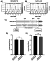
Growth and lentiviral yield performance was unaffected by nuclease-engineering of host cells. (A) HEK293TS and (B) NuPro-2S cell lines isolated in Figure 2 were cryopreserved and cryorevived using standard procedures, and their subsequent viable cell density (VCD) and viability (black and gray data points, respectively) were logged over six passages over 19 days postrevival. For both metrics, error bars are mean ± standard deviation (SD) of duplicate 20 mL shake flask cultures. (C) Both cell lines were used for lentivirus production in procedures that were matched except for addition of Benzonase to 50 units/mL concentration to HEK293TS cells 2 h prior to harvest and addition of tetracycline to 1 μg/mL concentration to NuPro-2S cells 24 h prior to harvest. (D) Lentivirus-containing growth media from both cell lines were used to transduce HEK293T target cells. and the resulting titers were plotted as transducing units/mL (TU/mL). Error bars are the combined mean ± SD of n = 3 biological repeats, which are 20 mL runs of lentivirus production by transient transfection, and n = 3 technical repeats, which are flow cytometric measurements on transduced cells of three individual wells. (E) Material tested in (D) was also used for RT-qPCR, and the resulting viral genomes/mL (vg/mL) were plotted. Error bars are the combined mean ± SD of n = 3 20 mL runs of lentivirus production and n = 2 RT-qPCR determinations. Differences in both TU/mL and vg/mL were not significant (ns) by unpaired Student’s t test.
Figure 4
Figure 4
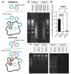
During lentivirus production, NuPro-2S cells give rise to medium-resident nuclease activity that reduces DNA impurity levels in situ. (A) Before and after the Figure 3C Benzonase addition, 10 mL samples of HEK293TS cells were centrifuged at 500 rcf for 5 min, and the supernatant was removed and retained as “Clarified Media”. Uncentrifuged samples were also taken and retained as “Whole Culture”. Equivalent NuPro2S samples were also taken. All samples were then incubated at 37 °C for a 1 h hold step. This diagram illustrates the anticipated presence of nuclease activity (red symbols) arising from NuPro-2S and the presence of lentivirus (green symbols) produced from both cell lines and confirmed in Figure 3. (B) “Clarified Media” samples (8 μL) were incubated with 2 μL of a 500 ng/μL 1 kb DNA ladder solution for 1 h prior to agarose gel electrophoresis. Samples were either pre- or posthold, as indicated, and taken from cultures of the indicated cell lines. Cultures had either had no additions or addition of tetracycline or Benzonase as detailed in Figure 3. (C) 40 μL samples, either pre- or posthold, from the indicated cultures were analyzed directly by agarose gel electrophoresis. (D) 100 μL serially diluted samples posthold from the indicated cultures were analyzed using the PicoGreen reagent, and the DNA was content plotted. Error bars represent the standard deviation of means arising from samples from n = 2 lentiviral production runs, each of which was used for n = 3 Pico Green-based determinations. Significance was determined by the unpaired Student’s t test. DNA concentration differences between unsupplemented HEK293TS and Benzonase-supplemented HEK293TS (p = 0.0921), and NuPro-2S (p < 0.0001) were significant (asterisks), while differences between Benzonase-supplemented HEK293TS and NuPro-2S were not (ns). All gel images are representative of duplicate gels used for analysis of duplicate samples.

Similar articles

Cited by

References

    1. July 24, 2020 Approval Letter - TECARTUS. U.S. Food and Drug Administration, July 24, 2020. https://www.fda.gov/media/140415/download (accessed 2022-12-21).
    1. August 30, 2017 Approval Letter - KYMRIAH. U.S. Food and Drug Administration, August 30, 2017. https://www.fda.gov/media/106989/download (accessed 2022-12-21).
    1. October 18, 2017 Approval Letter - YESCARTA. U.S. Food and Drug Administration, October 18, 2017. https://www.fda.gov/media/108458/download (accessed 2022-12-21).
    1. August 17, 2022 Approval Letter - ZYNTEGLO. U.S. Food and Drug Administration, August 17, 2022. https://www.fda.gov/media/160994/download (accessed 2022-12-21)
    1. September 16, 2022 Approval Letter - SKYSONA. U.S. Food and Drug Administration, September 16, 2022. https://www.fda.gov/media/161665/download (accessed 2022-12-21).

Publication types