. 2024 Apr:70:103038.
doi: 10.1016/j.redox.2024.103038. Epub 2024 Jan 11.

Endothelial H2S-AMPK dysfunction upregulates the angiocrine factor PAI-1 and contributes to lung fibrosis

Affiliations

Endothelial H2S-AMPK dysfunction upregulates the angiocrine factor PAI-1 and contributes to lung fibrosis

Xiangqi Chen et al. Redox Biol. 2024 Apr.

Abstract

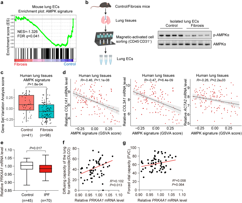
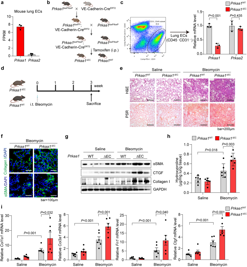
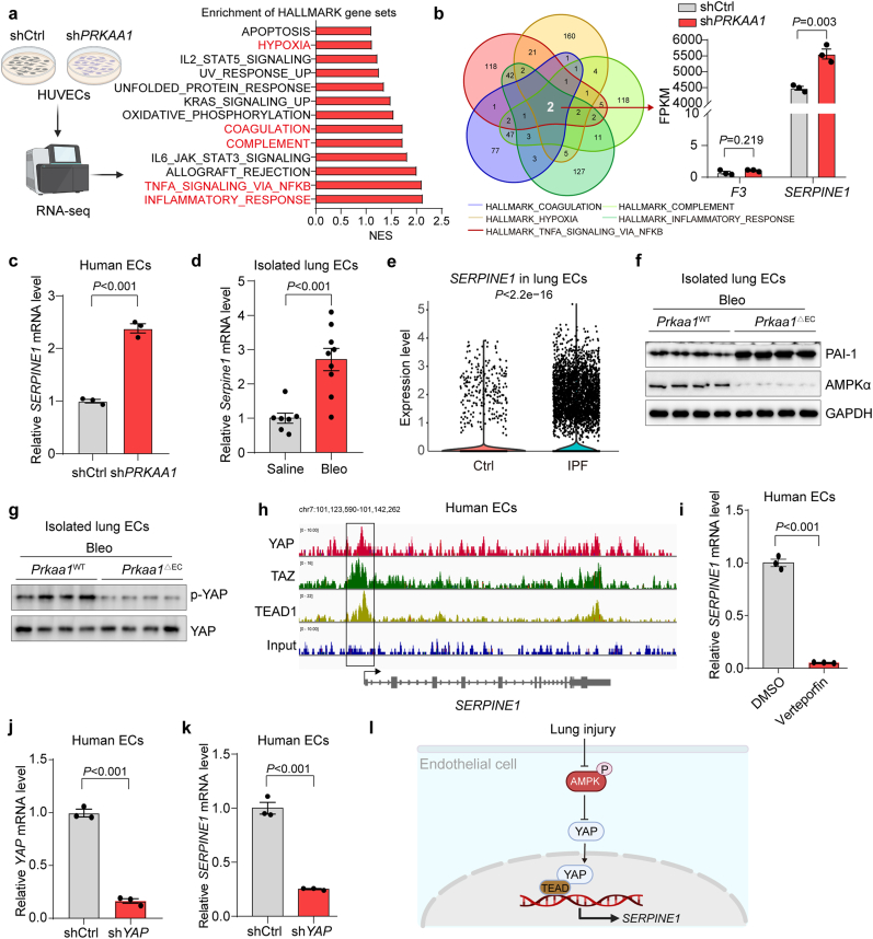
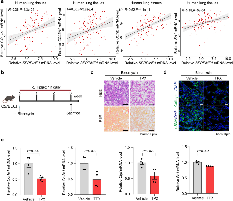
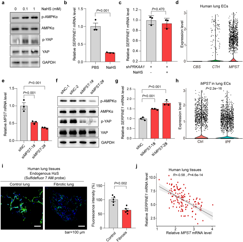
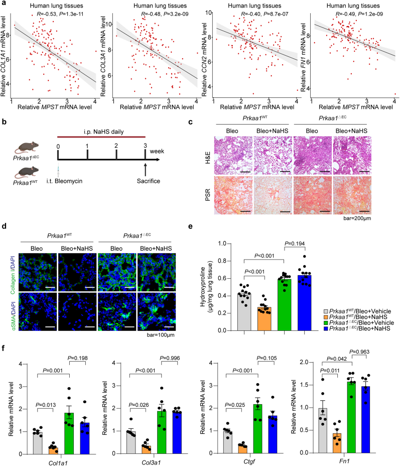
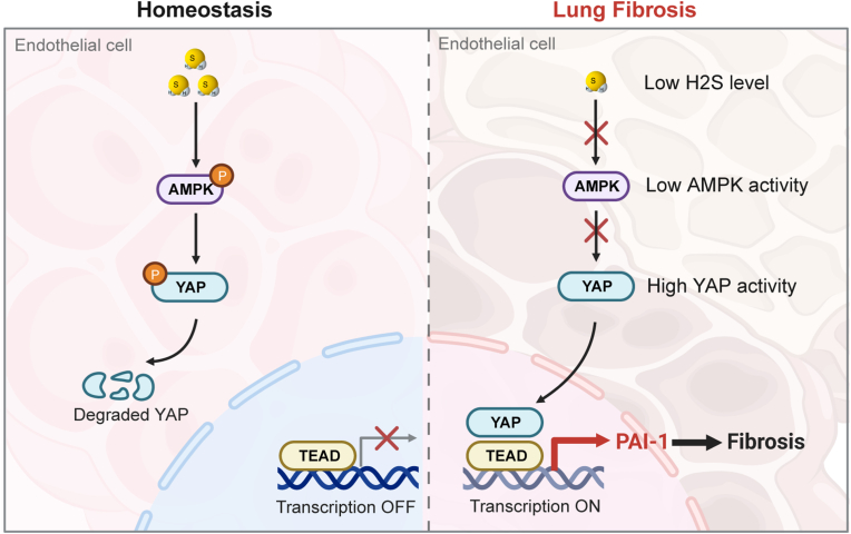
Dysfunction of the vascular angiocrine system is critically involved in regenerative defects and fibrosis of injured organs. Previous studies have identified various angiocrine factors and found that risk factors such as aging and metabolic disorders can disturb the vascular angiocrine system in fibrotic organs. One existing key gap is what sense the fibrotic risk to modulate the vascular angiocrine system in organ fibrosis. Here, using human and mouse data, we discovered that the metabolic pathway hydrogen sulfide (H2S)-AMP-activated protein kinase (AMPK) is a sensor of fibrotic stress and serves as a key mechanism upregulating the angiocrine factor plasminogen activator inhibitor-1 (PAI-1) in endothelial cells to participate in lung fibrosis. Activation of the metabolic sensor AMPK was inhibited in endothelial cells of fibrotic lungs, and AMPK inactivation was correlated with enriched fibrotic signature and reduced lung functions in humans. The inactivation of endothelial AMPK accelerated lung fibrosis in mice, while the activation of endothelial AMPK with metformin alleviated lung fibrosis. In fibrotic lungs, endothelial AMPK inactivation led to YAP activation and overexpression of the angiocrine factor PAI-1, which was positively correlated with the fibrotic signature in human fibrotic lungs and inhibition of PAI-1 with Tiplaxtinin mitigated lung fibrosis. Further study identified that the deficiency of the antioxidative gas metabolite H2S accounted for the inactivation of AMPK and activation of YAP-PAI-1 signaling in endothelial cells of fibrotic lungs. H2S deficiency was involved in human lung fibrosis and H2S supplement reversed mouse lung fibrosis in an endothelial AMPK-dependent manner. These findings provide new insight into the mechanism underlying the deregulation of the vascular angiocrine system in fibrotic organs.

Keywords: AMPK; H(2)S; Lung fibrosis; PAI-1; Vascular endothelial cells; YAP.


Conflict of interest statement

Declaration of competing interest The authors declare that they have no conflict of interest.

Figures

Fig. 1
Fig. 1
AMPK is inactivated in human and mouse fibrotic lungs. (a) The AMPK signaling pathway is repressed in endothelial cells (ECs) of mouse fibrotic lungs. Transcriptome data (GSE181508) of lung endothelial cells from bleomycin-induced fibrotic lungs and control lungs were subjected to Gene Set Enrichment Analysis (GSEA) of AMPK_SIGNALING_PATHWAY. (b) AMPK phosphorylation/activation is repassed in endothelial cells of mouse fibrotic lungs. Lung fibrosis was induced by bleomycin for three weeks, then endothelial cells from fibrotic and control lungs were isolated by Magnetic-Activated Cell Sorting (MACS) with indicated antibodies (CD45CD31+). Western blot was performed to test the phosphorylation/activation of AMPK in endothelial cells of fibrotic and control lungs. (c) The AMPK signaling pathway is repressed in human fibrotic lungs. Transcriptome data (GSE213001) of 41 control and 98 fibrotic lung samples were subjected to analyze the Gene Set Variation Analysis (GSVA) score of AMPK_SIGNALING_PATHWAY (AMPK signature). (d) AMPK signature is negatively correlated with the expression of fibrotic marker genes (COL1A1, COL3A1, ACTA2). Transcriptome data in (c) were subjected to Spearman correlation analysis. (e) AMPK mRNA (PRKAA1) is reduced in PBMCs from patients with lung fibrosis. PRKAA1 mRNA level from 45 control donors and 70 IPF patients PBMCs using the GEO public microarray dataset GSE38958. (f–g) PRKAA1 level is positively correlated with lung function parameters. The correlation of PRKAA1 mRNA level with lung function in patients with lung fibrosis was performed with Spearman correlation analysis using the GSE38958 microarray dataset. DLCO, diffusing capacity for carbon monoxide; FVC, forced vital capacity.
Fig. 2
Fig. 2
Endothelial cell-specific Prkaa1 knockout promotes lung fibrosis. (a) FPKM value of Prkaa1 and Prkaa2 in mouse lung ECs showing higher expression abundance of Prkaa1 (n = 3). Bulk RNA-seq data of mouse lung ECs was downloaded from the GEO database (GSE148893). (b) Schematic diagram for generating endothelial-specific Prkaa1 knockout mice. (c) Validation of Prkaa1 knockout efficacy in lung endothelial cells. Lung endothelial cells (CD45CD31+) were purified from Prkaa1WT and Prkaa1△EC mice with FACS, followed by qRT-PCR analysis of Prkaa1 and Prkaa2 mRNA levels (n = 3). (d) Schematic diagram showing mouse lung fibrosis model. Lung fibrosis was induced in Prkaa1WT and Prkaa1△EC mice by intratracheal injection with bleomycin. (e) H&E and PSR staining showing increased remodeling and collagen deposition in Prkaa1△EC mice compared with Prkaa1WT in bleomycin-induced fibrotic lungs. (f) Immunofluorescent staining of Collagen and alpha-smooth Muscle Actin (αSMA) showing increased extracellular matrix in Prkaa1△EC mice compared with Prkaa1WT. (g) Expression of fibrosis-related proteins (αSMA, CTGF, Collagen I) is increased in Prkaa1△EC mice lung tissue compared with Prkaa1WT. (h) Measurement of hydroxyproline content in lung tissues from Prkaa1△EC and Prkaa1WT mice (n = 6). (i) Measurement of fibrosis-related genes (Col1a1, Col3a1, Ctgf, Fn1) mRNA level in Prkaa1△EC and Prkaa1WT mice lung tissues (n = 6).
Fig. 3
Fig. 3
AMPK regulates PAI-1 expression through YAP in endothelial cells in fibrotic lungs. (a) AMPK regulates fibrosis-associated hallmarks in endothelial cells. PRKAA1 was silenced in HUVECs by lentivirus-mediated shRNA, followed by TGFβ treatment for 24 h. Then, HUVECs were collected for bulk RNA-seq, followed by gene set enrichment analysis (GSEA) of HALLMARK gene sets. The top enriched HALLMARK gene sets in endothelial cells with AMPK deficiency are shown. (b) SERPINE1 is a key downstream gene of AMPK in human endothelial cells. The intersection of key HALLMARK gene sets from a (red) identified F3 and SERPINE1 as two core genes in the key HALLMARK gene sets, and AMPK effects on the expression values (FPKM) of F3 and SERPINE1 are shown. (c) PRKAA1 knockdown increases SERPINE1 expression in human endothelial cells. HUVECs were treated as in (a), followed by qRT-PCR analysis of SERPINE1 mRNA level (n = 3). (d) Serpine1 expression is increased in endothelial cells from mouse fibrotic lungs. Mouse lung fibrosis was induced by bleomycin (Bleo), lung endothelial cells were purified with the MACS method and followed qRT-PCR analysis of Serpine1 expression in lung ECs from control (Saline) or fibrotic (Bleo) lungs (Saline, n = 7; Bleo, n = 9). (e) Violin plot showing increased SERPINE1 expression in lung ECs from human fibrotic lungs compared with control lungs. scRNA-seq data of human lung fibrosis (GSE159354) was downloaded from the GEO database, and the transcriptome of lung endothelial cells was analyzed. (f) Prkaa1 knockout upregulates Serpine1 (PAI-1) in endothelial cells from mouse fibrotic lungs. (g) Prkaa1 knockout activates YAP (reduced phosphorylation) in endothelial cells from mouse fibrotic lungs. (h) ChIP-Seq profiling of HUVECs showing YAP, TAZ, and TEAD1 binding to the promoter region of SERPINE1. GSE163458 ChIP-Seq data was downloaded from the GEO database. (i) Inhibition of YAP with verteporfin inhibits SERPINE1 expression in human endothelial cells. HUVECs were treated with YAP inhibitor verteporfin (0.25 μM) for 48 h, followed by qRT-PCR analysis of SERPINE1 (n = 3). (j–k) Knockdown of YAP reduces SERPINE1 expression in human endothelial cells. HUVECs were infected with lentivirus carrying shCtrl or shYAP for 48 h, followed by qRT-PCR analysis of YAP and SERPINE1 levels (n = 3). (l) Schematic diagram for AMPK-YAP-PAI-1 axis in endothelial cells of fibrotic lungs. (For interpretation of the references to colour in this figure legend, the reader is referred to the Web version of this article.)
Fig. 4
Fig. 4
PAI-1 participates in lung fibrosis in humans and mice. (a) SERPINE1 mRNA expression is positively correlated with fibrotic markers. Bulk RNA-seq data of 41 human control lung tissues and 98 fibrotic lung tissues (GSE213001) was used to analyze the correlation of SERPINE1 and the expression of fibrotic marker genes (COL1A1, COLA3A1, CCN2, FN1) with Spearman correlation analysis. (b) Design of PAI-1 inhibitor treatment of mouse lung fibrosis. Mouse lung fibrosis was induced by bleomycin, followed by PAI-1 inhibitor (Tiplaxtinin; 5 mg/kg/day; i.g.) for 21 days. (c) H&E and PSR staining reveal that Tiplaxtinin (TPX) reduces tissue remodeling and collagen deposition in fibrotic lungs. (d) Immunofluorescent staining reveals that Tiplaxtinin decreases Collagen and αSMA expression in fibrotic lungs. (e) qRT-PCR analysis reveals that Tiplaxtinin decreases the expression of fibrotic marker genes (Col1a1, Col3a1, Ctgf, Fn1) in fibrotic lungs (n = 4).
Fig. 5
Fig. 5
Hydrogen sulfide regulates the AMPK-YAP-PAI-1 axis in endothelial cells. (a) Hydrogen sulfide (H2S) donor NaHS induces AMPK activation and YAP phosphorylation/inactivation in human endothelial cells. HUVECs were treated with/without NaHS for 24 h, followed by Western blot analysis of AMPK and YAP activation. (b) H2S donor NaHS inhibits SERPINE1 expression in human endothelial cells. HUVEC cells were treated with NaHS (1 mM) for 24 h, followed by qRT-PCR analysis of SERPINE1 expression (n = 3). (c) PRKAA1 knockdown counteracts the inhibitory effect of H2S on SERPINE1 expression in human endothelial cells. HUVECs were infected with lentivirus carrying shPRKAA1 and treated with NaHS (1 mM) for an additional 24 h, followed by qRT-PCR analysis of SERPINE1 expression (n = 3). (d) Expression levels of the three H2S synthetases (CBS, CTH, MPST) in human lung ECs. Transcriptome data of human lung endothelial cells were obtained from the public dataset GSE159354. (e) siRNA-mediated knockdown of MPST in human endothelial cells. HUVECs were transfected with siMPST or siNC for 24 h, followed by qRT-PCR analysis of MPST mRNA level (n = 3). (f) Knockdown of MPST inhibits AMPK activation and YAP phosphorylation/inactivation in human endothelial cells. HUVECs were treated as in (e). (g) Knockdown of MPST increases the expression of SERPINE1 in human endothelial cells. HUVECs were treated as in (e) (n = 3). (h) Expression of the key H2S synthetase MPST is reduced in endothelial cells from human fibrotic lungs. The transcriptome of endothelial cells from fibrotic and control lung tissues was analyzed with the public scRNA-seq dataset GSE159354. (i) H2S level is reduced in human fibrotic lungs. Endogenous H2S in human fibrotic and control lungs were detected with the probe Sulfidefluor 7 AM (n = 5). (j) MPST expression is negatively correlated with SERPINE1 in human lung tissues. The Spearman correlation analysis of SERPINE1 mRNA expression level with MPST mRNA expression level in human lung tissues was performed using the public dataset GSE213001.
Fig. 6
Fig. 6
Hydrogen sulfide reverses lung fibrosis in an endothelial AMPK-dependent manner. (a) H2S synthetase MPST is negatively correlated with fibrotic markers in human fibrotic lungs. The Spearman correlation was performed to analyze the correlation of MPST mRNA expression level with fibrotic marker genes (COL1A1, COL3A1, CCN2, FN1) mRNA expression level in human lung tissues using the public dataset GSE213001. (b) Design of H2S-mediated treatment of lung fibrosis in WT mice and mice with endothelial Prkaa1 knockout. Lung fibrosis in WT mice and mice with endothelial Prkaa1 knockout was induced with bleomycin, followed by H2S donor NaHS (10 mg/kg/day) treatment for 21 days. (c) H&E and PSR staining reveal decreased tissue remodeling and collagen deposition in NaHS-treated mice, while this effect is abolished by endothelial-specific Prkaa1 knockout. (d) Immunofluorescent staining reveals decreased Collagen and αSMA expression in fibrotic lung tissues in NaHS-treated mice, while this effect is abolished by endothelial-specific Prkaa1 knockout. (e) Measurement of fibrotic marker hydroxyproline in lung tissues shows decreased fibrosis in NaHS-treated mice, while this effect is abolished by endothelial-specific Prkaa1 knockout (n = 12). (f) Fibrosis-related gene (Col1a1, Col3a1, Ctgf, Fn1) expression levels are decreased in NaHS-treated mice, while this effect is abolished by endothelial-specific Prkaa1 knockout (n = 6).
Fig. 7
Fig. 7
Endothelial H2S-AMPK regulates the YAP-PAI-1 axis to participate in lung fibrosis. During lung injury, loss of endogenous H2S triggers the inactivation of AMPK in endothelial cells. As a result, AMPK fails to induce YAP phosphorylation, leading to the translocation of YAP into the endothelial nucleus, thereby enhancing PAI-1 transcription and subsequently promoting the development of lung fibrosis.

Similar articles

Cited by

References

    1. Zhao X., Kwan J.Y.Y., Yip K., Liu P.P., Liu F.F. Targeting metabolic dysregulation for fibrosis therapy. Nat. Rev. Drug Discov. 2020;19:57–75. - PubMed
    1. Henderson N.C., Rieder F., Wynn T.A. Fibrosis: from mechanisms to medicines. Nature. 2020;587:555–566. - PMC - PubMed
    1. Koudstaal T., Funke-Chambour M., Kreuter M., Molyneaux P.L., Wijsenbeek M.S. Pulmonary fibrosis: from pathogenesis to clinical decision-making. Trends Mol. Med. 2023;29:1076–1087. - PubMed
    1. Liu G.Y., Budinger G.R.S., Dematte J.E. Advances in the management of idiopathic pulmonary fibrosis and progressive pulmonary fibrosis. BMJ. 2022;377 - PubMed
    1. Karampitsakos T., Juan-Guardela B.M., Tzouvelekis A., Herazo-Maya J.D. Precision medicine advances in idiopathic pulmonary fibrosis. EBioMedicine. 2023;95:104766. - PMC - PubMed

MeSH terms

Substances