. 2024 Feb:483:116833.
doi: 10.1016/j.taap.2024.116833. Epub 2024 Jan 23.

Prenatal arsenite exposure alters maternal cardiac remodeling during late pregnancy

Affiliations

Prenatal arsenite exposure alters maternal cardiac remodeling during late pregnancy

Nicole Taube et al. Toxicol Appl Pharmacol. 2024 Feb.

Abstract

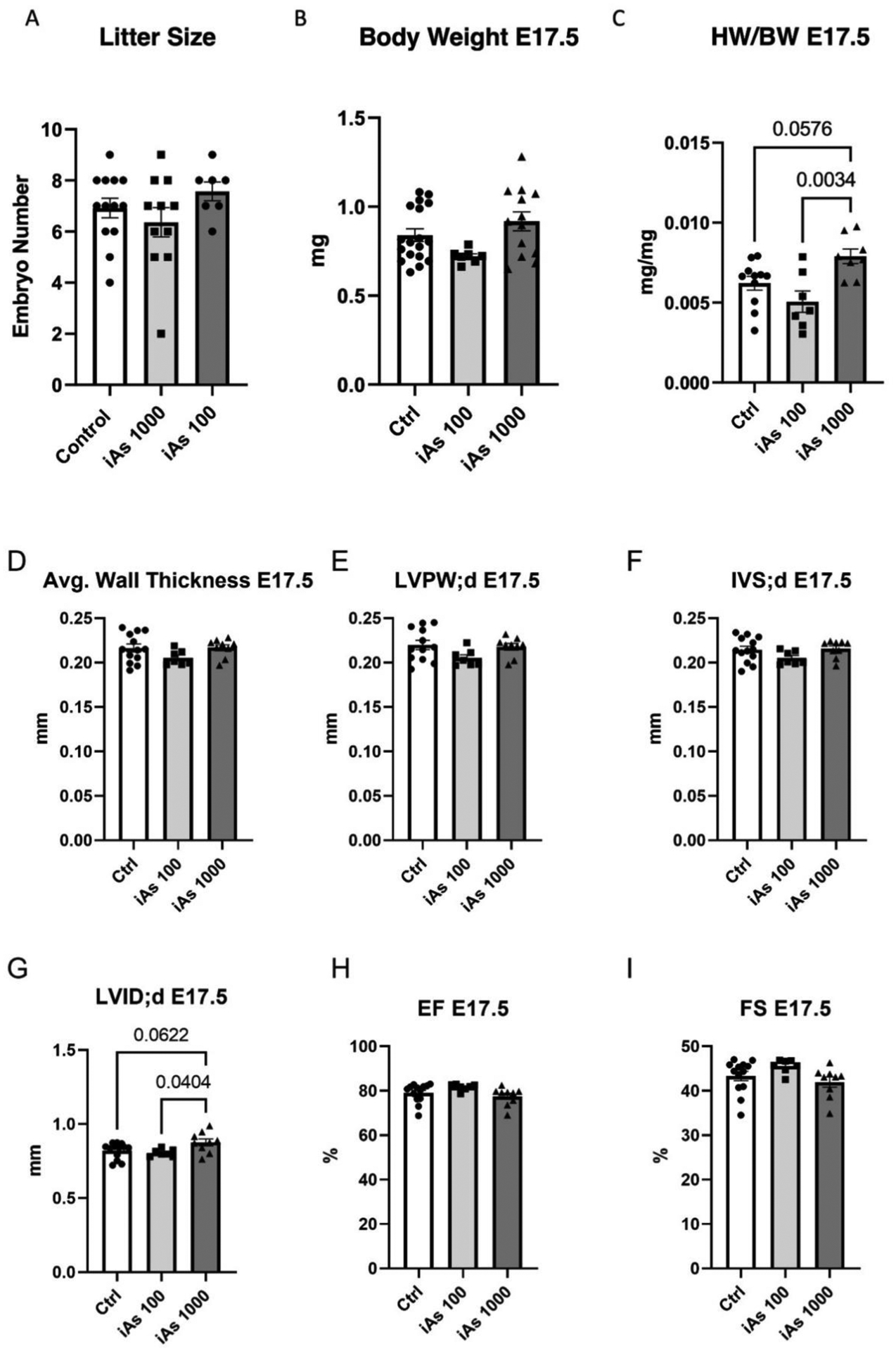
Exposure to inorganic arsenic through drinking water is widespread and has been linked to many chronic diseases, including cardiovascular disease. Arsenic exposure has been shown to alter hypertrophic signaling in the adult heart, as well as in utero offspring development. However, the effect of arsenic on maternal cardiac remodeling during pregnancy has not been studied. As such, there is a need to understand how environmental exposure contributes to adverse pregnancy-related cardiovascular events. This study seeks to understand the impact of trivalent inorganic arsenic exposure during gestation on maternal cardiac remodeling in late pregnancy, as well as offspring outcomes. C57BL/6 J mice were exposed to 0 (control), 100 or 1000 μg/L sodium arsenite (NaAsO2) beginning at embryonic day (E) 2.5 and continuing through E17.5. Maternal heart function and size were assessed via transthoracic echocardiography, gravimetric measurement, and histology. Transcript levels of hypertrophic markers were probed via qRT-PCR and confirmed by western blot. Offspring outcomes were assessed through echocardiography and gravimetric measurement. We found that maternal heart size was smaller and transcript levels of Esr1 (estrogen receptor alpha), Pgrmc1 (progesterone receptor membrane component 1) and Pgrmc2 (progesterone receptor membrane component 2) reduced during late pregnancy with exposure to 1000 μg/L iAs vs. non-exposed pregnant controls. Both 100 and 1000 μg/L iAs also reduced transcription of Nppa (atrial natriuretic peptide). Akt protein expression was also significantly reduced after 1000 μg/L iAs exposure in the maternal heart with no change in activating phosphorylation. This significant abrogation of maternal cardiac hypertrophy suggests that arsenic exposure during pregnancy can potentially contribute to cardiovascular disease. Taken together, our findings further underscore the importance of reducing arsenic exposure during pregnancy and indicate that more research is needed to assess the impact of arsenic and other environmental exposures on the maternal heart and adverse pregnancy events.

Keywords: Arsenic; Cardiotoxicity; Cardiovascular disease; Maternal exposure; Pregnancy.


Conflict of interest statement

Declaration of competing interest The authors declare the following financial interests/personal relationships which may be considered as potential competing interests. Mark Kohr reports financial support was provided by National Heart Lung and Blood Institute. Nicole Taube reports financial support was provided by National Institute of Environmental Health Sciences. Haley Garbus reports financial support was provided by National Heart Lung and Blood Institute. Mark Kohr reports a relationship with National Heart Lung and Blood Institute that includes: funding grants. If there are other authors, they declare that they have no known competing financial interests or personal relationships that could have appeared to influence the work reported in this paper.

Figures

Figure 1:
Figure 1:. Diagram of Experimental Timeline and Endpoints.
Experimental timeline of iAs exposure and experimental endpoints for dams and offspring. Created with Biorender.
Figure 2:
Figure 2:. iAs exposure increases maternal cardiac function during pregnancy.
Cardiac parameters as measured by transthoracic echocardiography in both pregnant and non-pregnant mice including: cardiac output (A), stroke volume (B), heart rate (C), ejection fraction (D), fractional shortening (E), end diastolic volume (EDV; F), end systolic volume (ESV; G), average wall thickness (H), left ventricular internal diameter in diastole (LVID;d; I), and left ventricular internal diameter in systole (LVID;s; J) (*= P < 0.05, ** = P< 0.01 vs. control; n=7–9 mice/group). Significance was determined by two-way ANOVA with Tukey’s multiple comparisons test.
Figure 3:
Figure 3:. iAs exposure blunts maternal cardiac hypertrophy.
Heart weight divided by tibia length in pregnant and non-pregnant females with 1000 μg/L (A) and 100 μg/L (B) iAs exposure compared to non-exposed controls (n=11–18 mice/group; *= P < 0.05, ** = P< 0.01 vs. control). Quantification of fibrosis (C) and representative images (D, scale bar 2mm) in pregnant mice exposed to 1000 μg/L iAs vs. Non-exposed pregnant controls (Ctrl). Quantification of cardiomyocyte cross-sectional area (E) and representative images of cardiomyocyte cross-section (F, scale bar 100 μm) taken from 4 sections per heart with 25 cardiomyocytes per section (n=5 hearts/group, 100 cardiomyocytes/heart; **= P<0.01 vs. control). Significance was determined by two-way ANOVA with Sidak’s multiple comparisons test or Mann-Whitney test.
Figure 4:
Figure 4:. iAs downregulates estrogen receptor alpha transcripts during late pregnancy.
Myocardial mRNA transcript levels of estrogen receptor alpha (ESR1; A), progesterone receptor membrane component 1 (Pgrmc1; B), progesterone receptor membrane component 2 (Pgrmc2; C), and prolactin receptor (Prlr; D) in pregnant females at E17.5 as measured via RT-qPCR (**= P<0.01 vs. control). Transcript levels were normalized to Gapdh mRNA expression, which did not change with iAs exposure. Outliers were determined by the ROUT method (Q=1%), and significance was determined by one-way ANOVA with Sidak’s multiple comparisons test or Mann-Whitney test (n=7–9 mice/group).
Figure 5:
Figure 5:. iAs exposure reduces Akt and ANP transcripts in the prepartum heart.
Myocardial mRNA transcript levels of protein kinase B (Akt; A), natriuretic peptide A (ANP; B), vascular endothelial growth factor (Vegfa; C), potassium voltage-gated channel 4.3 (Kv4.3; D), endothelial nitric oxide synthase (Nos3; E), natriuretic peptide B (Nppb; F), β-myosin heavy chain (Myh7; G), α-myosin heavy chain (Myh6; H), skeletal muscle α-actin (Acta1; I), (*=P<0.05, n=7–9 hearts/group). Outliers were determined by the ROUT method (Q=1%), and significance was determined by a one-way ANOVA with Sidak’s multiple comparisons test.
Figure 6:
Figure 6:. iAs exposure reduces protein levels of Akt in the prepartum heart.
Myocardial protein expression of phosphorylated Akt at the activating site, S437, normalized to total Akt (A) in dam hearts (P= 0.0776 vs. control, n= 5 hearts/group). Protein expression of total Akt (B) in dam hearts (***P=0.0005 vs. control). Protein expression of phosphorylated ERK1/2 at the activating sites, T202/Y204 (C), and of total ERK1/2 (D). Protein levels of each target were normalized to total transferred protein levels. Outliers were determined by the ROUT method (Q=1%), and significance was determined by a Mann-Whitney test.
Figure 7:
Figure 7:. Offspring from iAs exposed dams show increased heart weight and left ventricular internal diameter at E17.5.
Litter size (A), body weight (B), and embryonic heart weight normalized to body weight (C), in offspring exposed to 100 and 1000 μg/L iAs during gestation (n= 16–18 litters/group). Cardiac parameters measured in utero using echocardiography including average wall thickness (D), left ventricular posterior wall thickness (LVPW; E), interventricular septum thickness (IVS; F), left ventricular internal diameter (LVID; G), ejection fraction (EF; H), and fractional shortening (FS; I). Outliers were determined by the ROUT method (Q=1%), and significance was determined by one-way ANOVA with Sidak’s multiple comparisons test.
Figure 8:
Figure 8:. Offspring exposed to iAs in utero have reduced body weight at P12.
Body weight at P12 (A), body weight stratified by sex (B), heart weight relative to body weight (C), and heart weight relative to body weight stratified by sex (D) measured gravimetrically. Cardiac functional parameters measured via echocardiography including average wall thickness (E), interventricular septum thickness (IVS; F), left ventricular posterior wall thickness (LVPW; G), left ventricular internal diameter (LVID; H), ejection fraction (EF; I), and fractional shortening (FS; J). Significance was determined by one-way ANOVA with Sidak’s multiple comparisons test.

Update of

References

    1. Substance Priority List | ATSDR. https://www.atsdr.cdc.gov/spl/index.html.
    1. Nordstrom DK PUBLIC HEALTH: Enhanced: Worldwide Occurrences of Arsenic in Ground Water. Science (1979) 296, 2143–2145 (2002). - PubMed
    1. Abernathy CO, Thomas DJ & Calderon RL Toxicity and Risk Assessment of Trace Elements Health Effects and Risk Assessment of Arsenic 1,2.
    1. Vahter M Mechanisms of arsenic biotransformation. Toxicology 181–182, 211–217 (2002). - PubMed
    1. Ferreccio C et al. Lung cancer and arsenic concentrations in drinking water in Chile. Epidemiology 11, 673–679 (2000). - PubMed