. 2024 Mar 16;86(3):300-307.
doi: 10.1292/jvms.23-0492. Epub 2024 Jan 24.

Establishment of an organ culture system to maintain the structure of mouse Müllerian ducts during development

Affiliations

Establishment of an organ culture system to maintain the structure of mouse Müllerian ducts during development

Shiori Kato et al. J Vet Med Sci. .

Abstract

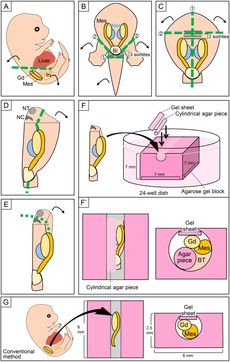
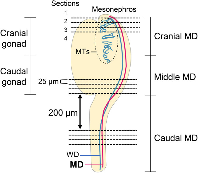
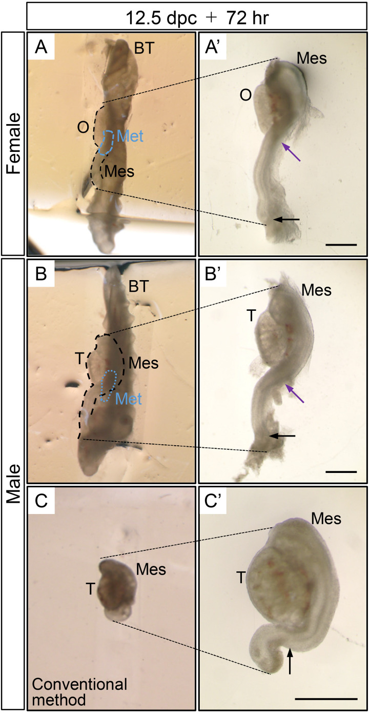
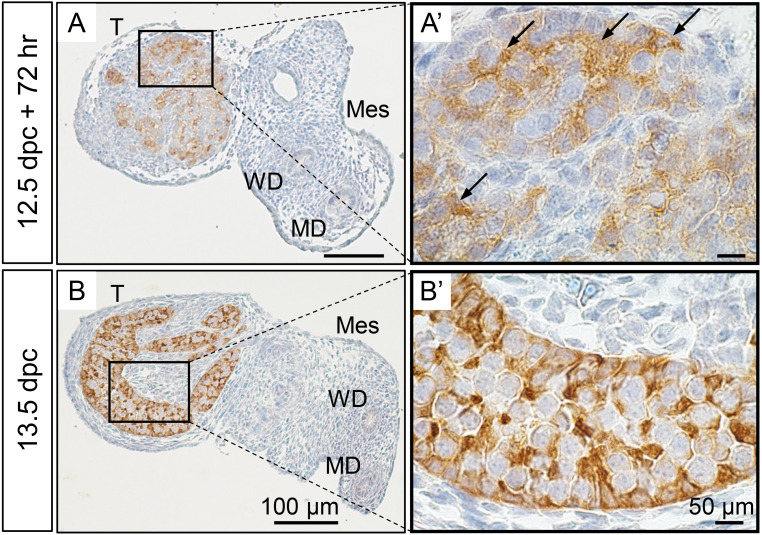
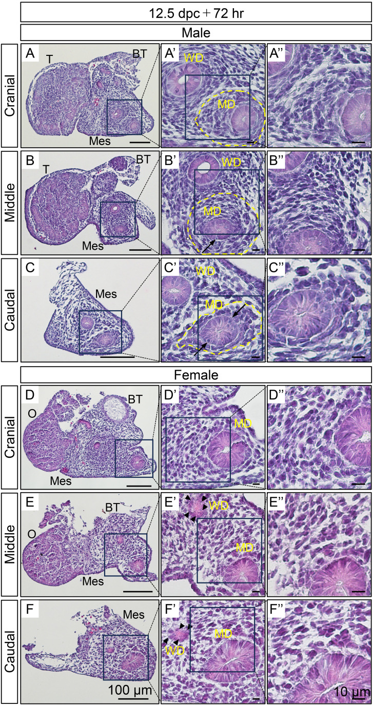
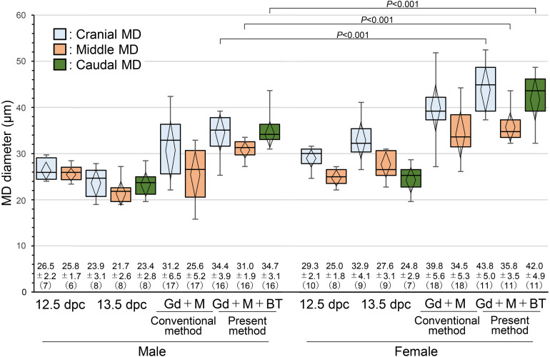
We previously showed that the anti-Müllerian hormone (AMH), infiltrating from the testis to the mesonephros reaches the cranial and middle regions of the Müllerian duct (MD) and induces their regression using an organ culture in mice. However, it is difficult to maintain structural integrity, such as the length and diameter and normal direction of elongation of the caudal region of the MD, in conventional organ culture systems. Therefore, the pathway of AMH to the caudal MD region remains uncharted. In this study, we established an organ culture method that can maintain the morphology of the caudal region of the MD. The gonad-mesonephros complex, metanephros, and urinary bladder of mouse fetuses at 12.5 dpc attached to the body trunk were cultured on agarose gels for 72 hr. The cultured caudal region of the mesonephros was elongated along the body trunk, and the course of the mesonephros was maintained in many individuals. In males, mesenchymal cells aggregated around the MD after culture. Moreover, the male MD diameter was significantly smaller than the female. Based on these results, it was concluded that the development of the MD was maintained in the present organ culture system. Using this culture system, AMH infiltration to the caudal region of the MD can be examined without the influence of AMH in the blood. This culture system is useful for clarifying the regression mechanism of the caudal region of the MD.

Keywords: Müllerian duct regression; anti-Müllerian hormone (AMH); organ culture; secretion manner; sexual differentiation.


Conflict of interest statement

The authors have no conflicts of interest.

Figures

Fig. 1.
Fig. 1.
Schematic diagram of the dissection and culture procedures. (A) The cranial side above the liver and the intestine (In) were removed from a mouse fetus at 12.5 dpc. (B) The tail was resected at the 3 somites caudally from the base of the tail, and both hindlimbs were also resected. (C) The body trunk (BT) was split into right and left halves at the midline, and the cranial side of the body was resected at a position 2 somites cranially from the most cranial part of the gonads (Gd). (D) The body wall outside the mesonephros (Mes) was resected. (E) The region containing the neural tube (NT) was resected. (F and F’) The reshaped culture piece was placed in a groove of agarose gel block with the dorsal side facing down. A cylindrical agar piece was placed on the medial side of the culture piece. A gel sheet was placed over the culture piece. (G) Conventional culture method. The gonad–mesonephros complex was extracted from the fetus and placed in a groove of agarose gel block. A gel sheet was placed over the gonad–mesonephros complex [9]. Bl, bladder; Met, metanephros; NC, notochord.
Fig. 2.
Fig. 2.
Measurement position of the Müllerian duct (MD) diameter. The short diameters of the cranial, middle, and caudal MD were measured at intervals of 25 μm for four HE-stained sections. MTs, mesonephric tubules; WD, Wolffian duct.
Fig. 3.
Fig. 3.
Gonad–mesonephros (Mes) complex and body trunk (BT) after 72 hr of culture. (A) An ovary (O)–mesonephros complex and body trunk after culture. The body trunk extended straight within the groove of the agarose gel block. The mesonephros was elongated along the body trunk. (A’) The ovary–mesonephros complex was excised from the body trunk after culture. The mesonephros extended along the ovary, changed direction at the caudal end of the ovary (purple arrow), and continued its extension along the body trunk. (B) A testis (T)–mesonephros complex and body trunk after culture. The body trunk extended straight within the groove of the agarose gel block. The mesonephros was elongated along the body trunk. (B’) The testis–mesonephros complex excised from the body trunk after culture. The mesonephros extended along the testis, changed direction at the caudal end of the testis (purple arrow), and continued its extension along the body trunk. (C and C’) A testis–mesonephros complex cultured by the previous method, i.e., with the gonad–mesonephros complex alone [9]. The mesonephros of the caudal region not attached to the testis was strongly curved. Testis cord-like structures were clearly observed in the testis. Black arrows indicate the end of the Müllerian duct. Met, Metanephros.
Fig. 4.
Fig. 4.
Immunoreactivity of anti-Müllerian hormone (AMH) in sections of the testis (T)–mesonephros (Mes) complexes after 72 hr of culture and at 13.5 dpc. (A) Immunoreactivities for AMH were detected within the cultured testis cord, and strong AMH immunoreactivities were also observed (A’; arrows). (B) Immunoreactivities for AMH were detected within the testis cord at 13.5 dpc. Sertoli cells showed strong immunoreactivity for AMH (B’). MD, Müllerian duct; WD, Wolffian duct.
Fig. 5.
Fig. 5.
HE-stained sections of male (A–C) and female (D–F) gonad–mesonephros (Mes) complexes after 72 hr of culture and enlarged images focusing on the reproductive tracts (A’–F’) and mesenchymal cells around the Müllerian duct (MD; A”–F”). In males (A’–C’ and A”–C”), lumens were formed in the cranial, middle, and caudal MD. The mesenchymal cells around the cranial, middle, and caudal MD were aggregated (yellow dotted lines), and degenerating cells (arrows) were also observed. The lumen of the Wolffian duct (WD) was clearly formed. In females (D’–F’ and D”–F”), lumens were formed in the cranial, middle, and caudal MD. Mesenchymal cells around the MD were not aggregated. Numerous vacuoles (arrowheads) were observed in the WD epithelium. BT, body trunk; O, ovary; T, testis.
Fig. 6.
Fig. 6.
The diameters of the cranial, middle, and caudal Müllerian ducts (MD) at 12.5 dpc, 13.5 dpc, and after 72 hr of culture with the gonad (Gd)–mesonephros (M) complex alone and with gonad–mesonephros complex attached to the body trunk (BT). The male and female caudal MD diameter at 12.5 dpc could not be measured because the MD had not extended to the caudal region. The male and female caudal MD cultured with the gonad–mesonephros complex alone could not be measured because the morphology of the caudal MD was not maintained. In males, the diameter of the middle MD was smaller than that of the cranial MD at 12.5 dpc, 13.5 dpc, and after 72 hr of culture by either culture method. At 13.5 dpc, the diameter of the caudal MD was larger than that of the middle MD. The caudal MD diameter was also larger than the middle MD diameter when the gonad–mesonephros complex was cultured attached to the body trunk. In females, the middle MD diameter was smaller than the cranial MD diameter at 12.5 dpc, 13.5 dpc, and after 72 hr of culture by either culture method. At 13.5 dpc, the diameter of the caudal MD was smaller than that of the middle MD. On the other hand, the caudal MD diameter was larger than the middle MD diameter when the complex was cultured attached to the body trunk. The diameters of the male cranial, middle, and caudal MD were significantly smaller than those of females (P<0.001). The rhombus in the boxplot indicates the mean ± SD. Data are presented as means ± SD. Numbers in parentheses are sample numbers.

Similar articles

References

    1. Arango NA, Kobayashi A, Wang Y, Jamin SP, Lee HH, Orvis GD, Behringer RR. 2008. A mesenchymal perspective of Müllerian duct differentiation and regression in Amhr2-lacZ mice. Mol Reprod Dev 75: 1154–1162. doi: 10.1002/mrd.20858 - DOI - PubMed
    1. Bentvelsen FM, Brinkmann AO, van der Schoot P, van der Linden JE, van der Kwast TH, Boersma WJ, Schröder FH, Nijman JM. 1995. Developmental pattern and regulation by androgens of androgen receptor expression in the urogenital tract of the rat. Mol Cell Endocrinol 113: 245–253. doi: 10.1016/0303-7207(95)03593-V - DOI - PubMed
    1. Donahoe PK, Ito Y, Hendren WH., 3rd. 1977. A graded organ culture assay for the detection of Mullerian inhibiting substance. J Surg Res 23: 141–148. doi: 10.1016/0022-4804(77)90202-5 - DOI - PubMed
    1. Donahoe PK, Ito Y, Marfatia S, Hendren WH., 3rd. 1976. The production of Mullerian inhibiting substance by the fetal, neonatal and adult rat. Biol Reprod 15: 329–334. doi: 10.1095/biolreprod15.3.329 - DOI - PubMed
    1. Dyche WJ. 1979. A comparative study of the differentiation and involution of the Mullerian duct and Wolffian duct in the male and female fetal mouse. J Morphol 162: 175–209. doi: 10.1002/jmor.1051620203 - DOI - PubMed

Substances

Supplementary concepts