. 2024 Jan 24;14(1):2048.
doi: 10.1038/s41598-024-52352-5.

Yeast 26S proteasome nuclear import is coupled to nucleus-specific degradation of the karyopherin adaptor protein Sts1

Affiliations

Yeast 26S proteasome nuclear import is coupled to nucleus-specific degradation of the karyopherin adaptor protein Sts1

Carolyn Allain Breckel et al. Sci Rep. .

Abstract

In eukaryotes, the ubiquitin-proteasome system is an essential pathway for protein degradation and cellular homeostasis. 26S proteasomes concentrate in the nucleus of budding yeast Saccharomyces cerevisiae due to the essential import adaptor protein Sts1 and the karyopherin-α protein Srp1. Here, we show that Sts1 facilitates proteasome nuclear import by recruiting proteasomes to the karyopherin-α/β heterodimer. Following nuclear transport, the karyopherin proteins are likely separated from Sts1 through interaction with RanGTP in the nucleus. RanGTP-induced release of Sts1 from the karyopherin proteins initiates Sts1 proteasomal degradation in vitro. Sts1 undergoes karyopherin-mediated nuclear import in the absence of proteasome interaction, but Sts1 degradation in vivo is only observed when proteasomes successfully localize to the nucleus. Sts1 appears to function as a proteasome import factor during exponential growth only, as it is not found in proteasome storage granules (PSGs) during prolonged glucose starvation, nor does it appear to contribute to the rapid nuclear reimport of proteasomes following glucose refeeding and PSG dissipation. We propose that Sts1 acts as a single-turnover proteasome nuclear import factor by recruiting karyopherins for transport and undergoing subsequent RanGTP-initiated ubiquitin-independent proteasomal degradation in the nucleus.


Conflict of interest statement

No competing interests declared.

Figures
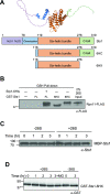
Figure 1
Figure 1
The Sts1 six-helix bundle is sufficient for proteasome interaction, but Sts1 degradation requires its N-terminus. (A) Predicted structure from AlphaFold2, (top), and predicted domain architecture of Sts1 and the likely functional elements based on protein sequence analysis and comparison with the crystal structure of the S. pombe homolog Cut8 (bottom). “NLS1” and “NLS2” indicate the two basic segments of the bipartite nuclear localization signal. Both the N-terminus and C-terminus are likely to be unstructured. Truncation mutants Sts1(116–276) and Sts1(116–319) are represented as “6HX” and “6HC,” respectively. (B) The Sts1 six-helix bundle is sufficient for interaction with 26S proteasomes. Purified recombinant species of GST-Sts1, GST-Sts1/Srp1-6His, GST-Sts1(116–276), or GST-Sts1(116–319) were immobilized on a glutathione (GSH) resin and incubated with 26S proteasomes purified from yeast to detect interactions. “FL” indicates full-length GST-Sts1. 26S proteasome input represents 2% of incubated proteasomes. The migration of Rpn11-FLAG is distorted by the closely migrating 6HX GST fusion. (C) Sts1 degradation is not observed when the Sts1 N-terminus is blocked. In vitro degradation analysis of purified recombinant MBP-Sts1 by 26S proteasomes purified from yeast. Degradation was measured at room temperature. (D) The Sts1 six-helix bundle (when fused to GST) is not sufficient to initiate its proteasomal degradation in vitro. Degradation assay as in (C) conducted using purified recombinant GST-Sts1(116–276). For “3 + MG” sample, proteasomes were incubated with 50 μM MG132 proteasome inhibitor for 10 min prior to addition of Sts1. Images have been cropped for clarity and original blots are presented in Supplementary Fig. 5.
Figure 2
Figure 2
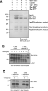
Sts1 proteasomal degradation correlates with removal of Kap95 by RanGTP. (A) Sts1 forms a ternary complex with Srp1 and Kap95 that can be selectively disrupted by the addition of RanGTP. Recombinant GST-Sts1 was immobilized on GSH resin and incubated with purified Kap95 and recombinant Srp1-6His to form a ternary complex. The complex was incubated with recombinant 6His-Gsp1GDP (RanGDP) or 6His-Gsp1GTP (RanGTP) to detect interactions and disassembly of the Sts1/Srp1/Kap95 complex. Pull-downs conducted at 4 °C. (B) Degradation of Sts1 is initiated when RanGTP removes karyopherin proteins from the Sts1 N-terminus. In vitro degradation assay as in Fig. 1C using the purified complex of recombinant Sts1-6His/GST-Srp1/Kap95 incubated with 26S proteasomes purified from yeast. After 3 h, purified recombinant RanGTP was added to the reaction mixture. For “6 + MG” sample, proteasomes were incubated with 50 μM MG132 proteasome inhibitor for 10 min prior to addition of the Sts1 complex. (C) Neither RanGTP nor RanGDP causes Sts1 instability in the absence of proteasomes. In vitro degradation assay as in (B), using the purified complex of Sts1-6His/GST-Srp1/Kap95 in the presence of purified RanGTP and RanGDP, respectively (as in A). Images have been cropped for clarity and original blots and gels are presented in Supplemental Fig. 5.
Figure 3
Figure 3
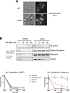
Sts1 proteasomal degradation occurs in the cell nucleus following nuclear import. (A) Sts1 accumulates in the cell nucleus when proteasomes are catalytically inactive. Wild-type and catalytically inactive proteasome mutant cim3-1 yeast expressing the plasmid pRS415-GPD-Sts1-GFP were visualized by fluorescence microscopy at the restrictive temperature for cim3-1. Scale bar, 5 μm. (B) Sts1 degradation occurs when proteasomes are sequestered inside the nucleus but not when sequestered at the plasma membrane or ribosome. Using the Anchor Away yeast system, 26S proteasomes were anchored to the plasma membrane, ribosome, or chromatin. Each anchoring strain, as well as a control strain with no proteasome anchor, also bears chromosomally tagged STS1-3xFLAG. Cycloheximide-chase analysis was performed to determine the degradation rates of Sts1-3xFLAG in the presence (+ Rapa) or absence (+ DMSO) of proteasome sequestration in different cellular compartments. Cells were grown at 30 °C, treated with 10 μg/mL of either rapamycin or DMSO for two hours, and cycloheximide was added at time 0 to block further protein synthesis. FLAG immunoprecipitation was performed on cell extracts to enrich for Sts1-3xFLAG. Bottom panels: quantification of cycloheximide-chase data from at least three experiments in the presence (right) or absence (left) of proteasome sequestration. Images have been cropped for clarity and original blots are presented in Supplemental Fig. 5.
Figure 4
Figure 4
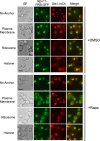
Sts1 can undergo nuclear import without binding proteasomes and accumulates in the nucleus even with proteasomes sequestered in the cytoplasm. The Anchor Away strains used in Fig. 3B were transformed with pRS415-MET25-Sts1-mCherry-FLAG plasmid to visualize Sts1 localization during proteasome sequestration (via chromosomally tagged RPN11-FRB-GFP) by fluorescence microscopy after 3 h of rapamycin or DMSO treatment. Cells were grown and treated with either DMSO or rapamycin as in Fig. 3B. Scale bar, 5 μm. Images have been false-colored and cropped for clarity.
Figure 5
Figure 5
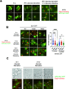
Sts1 not detected in proteasome storage granules (PSGs) or P-bodies under glucose starvation and does not participate in reimport of proteasomes from PSGs. (A) Sts1 does not localize to PSGs during glucose starvation and is not found in nuclei following reimport of proteasomes from PSGs. Yeast bearing the sts1∆ mutation and chromosomal RPN2-mCherry were transformed with plasmid pRS415-GPD-Sts1-GFP for fluorescence microscopy. Cells were grown in rich medium (2% glucose) at 30 °C and imaged (0 h of glucose starvation). Cells harvested and subsequently grown in low-glucose medium (0.025% glucose) for 2 days and imaged, then supplemented with 2% glucose and imaged after 1 h of glucose refeeding. (B) Import-defective Sts1 mutants do not exhibit a proteasome localization defect following reimport of proteasomes from PSGs. Yeast described in panel A were transformed with plasmids pRS415-GPD-Sts1-GFP, pRS415-GPD-Sts1(R38D)-GFP, or pRS415-GPD-Sts1(R65D)-GFP for fluorescence microscopy. Cells were treated as in panel A. For quantification, at least 100 cells were counted from three replicates to determine the nucleus to cytoplasm ratio (N/C ratio) of Rpn2-mCherry in cells (right panel). A t-test was used to determine the statistical significance of differences in localization (****p < 0.0001, “ns” indicates no significant difference). (C) Sts1 does not localize to P-bodies upon glucose starvation. Yeast bearing chromosomally tagged DCP2-mCherry were transformed with plasmid pRS415-GPD-Sts1-GFP for fluorescence microscopy. Cells were grown in rich media and transferred to low-glucose media as in panel A for three days and imaged. Scale bars, 5 μm. Arrowheads indicate either PSGs (A and B) or P-bodies (C). “V” marks the cell vacuole. “N” marks the cell nucleus. Images have been false-colored and cropped for clarity.
Figure 6
Figure 6
Model of Sts1/karyopherin-α/β-mediated nuclear import of 26S proteasomes and subsequent Sts1 ubiquitin-independent degradation. In the cytoplasm, Sts1, likely a homodimer, binds to karyopherin-α via interaction with the bipartite NLS sequence at the Sts1 N-terminus. Karyopherin-α recruits karyopherin-β and Sts1 subsequently binds to the 26S proteasome. This complex is imported into the nucleus via the nuclear pore complex. In the nucleus, RanGTP promotes removal of the karyopherin proteins from the Sts1 N-terminus. Once available, the unstructured Sts1 N-terminus is threaded into the proteasome RP and translocated into the CP to initiate ubiquitin-independent degradation of Sts1. This single turnover Sts1-mediated import mechanism occurs only during proliferative yeast growth.

Similar articles

Cited by

References

    1. Varshavsky A. The ubiquitin system, an immense realm. Annu. Rev. Biochem. 2012;81:167–176. doi: 10.1146/annurev-biochem-051910-094049. - DOI - PubMed
    1. Budenholzer L, Cheng CL, Li Y, Hochstrasser M. Proteasome structure and assembly. J. Mol. Biol. 2017;429:3500–3524. doi: 10.1016/j.jmb.2017.05.027. - DOI - PMC - PubMed
    1. Sahu I, Glickman MH. Proteasome in action: Substrate degradation by the 26S proteasome. Biochem. Soc. Trans. 2021;49:629–644. doi: 10.1042/BST20200382. - DOI - PMC - PubMed
    1. Pack C-G, et al. Quantitative live-cell imaging reveals spatio-temporal dynamics and cytoplasmic assembly of the 26S proteasome. Nat. Commun. 2014;5:3396. doi: 10.1038/ncomms4396. - DOI - PubMed
    1. Gödderz D, Schäfer E, Palanimurugan R, Dohmen RJ. The N-Terminal unstructured domain of yeast ODC functions as a transplantable and replaceable ubiquitin-independent degron. J. Mol. Biol. 2011;407:354–367. doi: 10.1016/j.jmb.2011.01.051. - DOI - PubMed