. 2024 May;31(5):801-809.
doi: 10.1038/s41594-023-01205-2. Epub 2024 Jan 24.

Cortactin stabilizes actin branches by bridging activated Arp2/3 to its nucleated actin filament

Affiliations

Cortactin stabilizes actin branches by bridging activated Arp2/3 to its nucleated actin filament

Tianyang Liu et al. Nat Struct Mol Biol. 2024 May.

Abstract

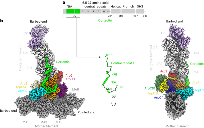
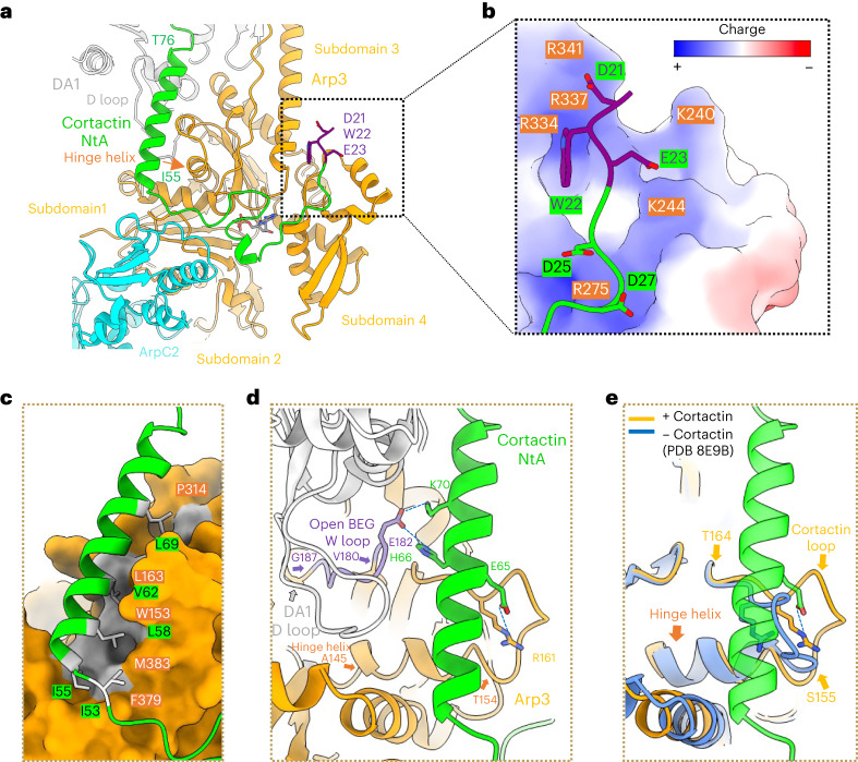
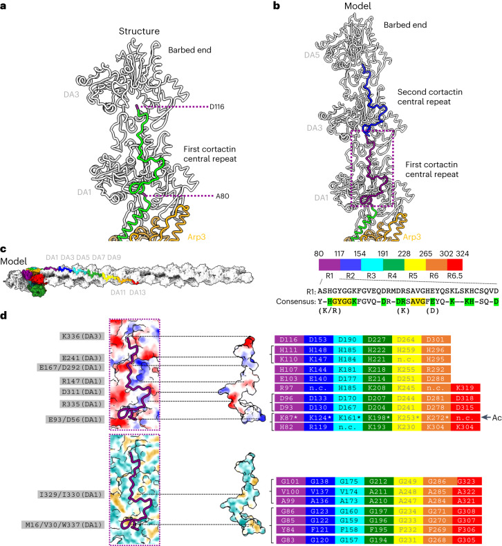
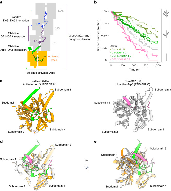
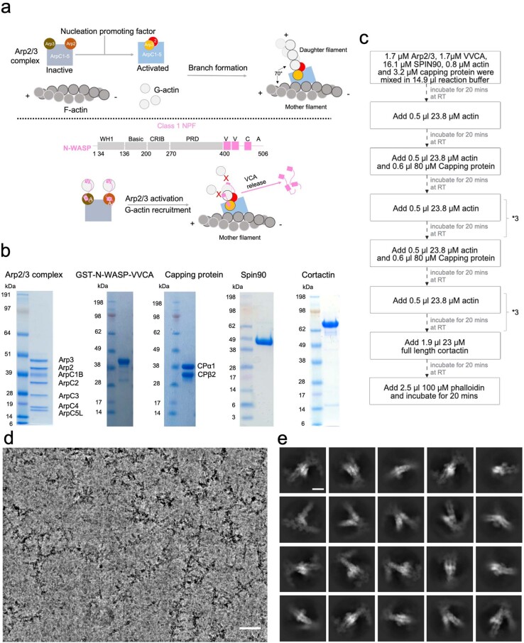
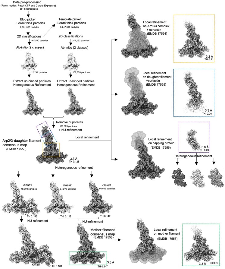
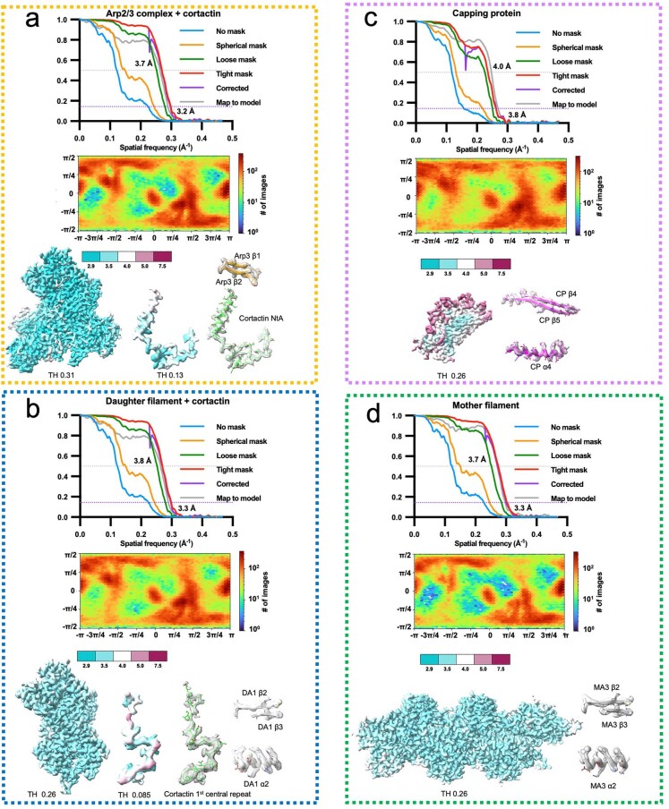
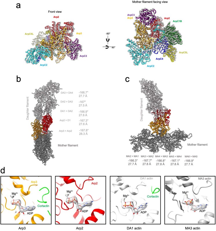
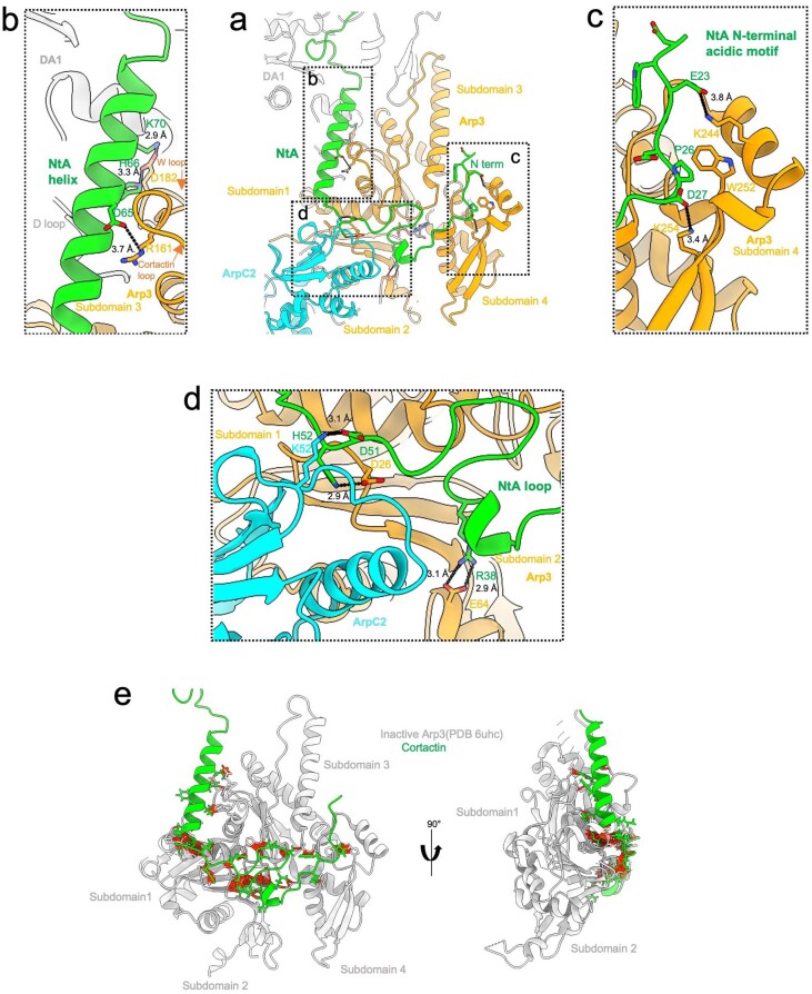
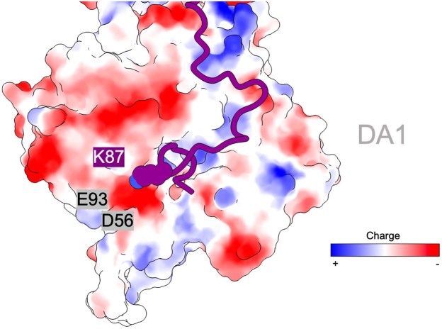
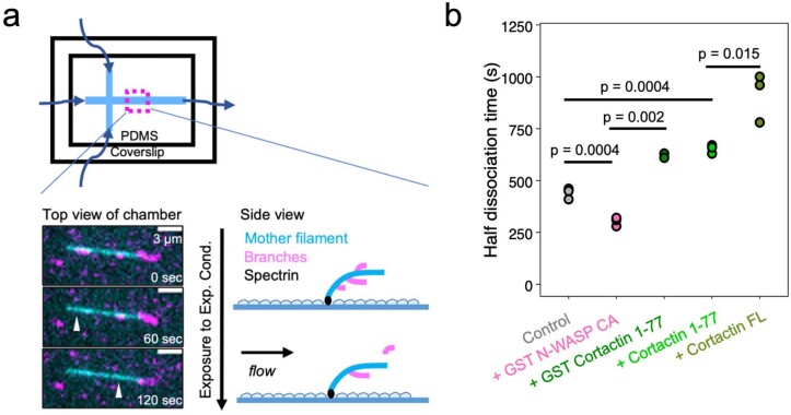
Regulation of the assembly and turnover of branched actin filament networks nucleated by the Arp2/3 complex is essential during many cellular processes, including cell migration and membrane trafficking. Cortactin is important for actin branch stabilization, but the mechanism by which this occurs is unclear. Given this, we determined the structure of vertebrate cortactin-stabilized Arp2/3 actin branches using cryogenic electron microscopy. We find that cortactin interacts with the new daughter filament nucleated by the Arp2/3 complex at the branch site, rather than the initial mother actin filament. Cortactin preferentially binds activated Arp3. It also stabilizes the F-actin-like interface of activated Arp3 with the first actin subunit of the new filament, and its central repeats extend along successive daughter-filament subunits. The preference of cortactin for activated Arp3 explains its retention at the actin branch and accounts for its synergy with other nucleation-promoting factors in regulating branched actin network dynamics.


Conflict of interest statement

The authors declare no competing interests.

Figures

Fig. 1
Fig. 1. Cortactin binds the daughter filament at Arp2/3-mediated actin branches.
a, Cortactin domain organization. b, Overview of the composite cryo-EM reconstruction of the cortactin-stabilized Arp2/3 actin branch, assembled from four local refined reconstructions, as shown in Extended Data Figs. 2 and 3. Densities of individual proteins are colored according to the labels, and the mother- and daughter-filament subunits are colored dark and light gray, respectively, and are labeled MA1, MA3, MA5 and MA6 and DA1–DA4. The free barbed and pointed ends of the mother and daughter filaments are also labeled. The central inset shows the cortactin model calculated from the cryo-EM reconstruction, with the visualized regions of the full-length protein mapped on to the cortactin schematic in a, as indicated.
Fig. 2
Fig. 2. Cortactin NtA binds to activated Arp3.
a, Overview of cortactin NtA (green and purple) and its interactions with Arp3 (orange) and ArpC2 (cyan). DWE motif (D21-W22-E23) residues are colored purple and shown as a stick model; the rest of cortactin NtA is depicted as a ribbon model. Cortactin residues D21–N54 meander across the Arp3 surface, and residues I55–T76 form an amphipathic α-helix. Subdomains in Arp3 are labeled, and the Arp3 hinge helix at the junction of subdomains 1 and 3 is indicated with an orange arrow. DA1 is the first subunit of the daughter filament, which, through the D-loop in its subdomain 2, forms longitudinal contacts with Arp3. Detailed views of interactions are shown in Extended Data Fig. 5a–d. b, Electrostatic interaction of the negatively charged cortactin N-terminal region that inserts into a positively charged pocket of Arp3. Arp3 is depicted in surface representation; acidic regions are shown in red, and basic regions are shown in blue with individual basic residues colored orange. Cortactin is depicted in a ribbon model, the DWE motif is shown as a purple stick model and residues 24–29 are colored green with acidic residues shown as a stick model. c, The hydrophobic interaction of the cortactin NtA α-helix (green ribbon model), with a hydrophobic groove on the surface of Arp3 (orange space-filling representation, with hydrophobic regions in gray). Residues on the interaction surface of cortactin and Arp3 are labeled in green and orange, respectively. d, The cortactin NtA α-helix stabilizes the activated Arp3 W-loop (residues 180–187, purple) conformation to enable DA1 actin subunit binding through its D-loop in the open barbed-end groove (BEG) of Arp3. Residues forming salt bridges are shown in stick representation. The distances between interacting residues are provided in Extended Data Fig. 5a–d. e, The cortactin NtA α-helix (green) stabilizes and interacts with a specific conformation of the Arp3 loop (residues 155–164), which we term the cortactin loop (in orange). The loop adopts a distinct conformation in the presence of cortactin, and its conformation is different in activated Arp2/3 in the absence of cortactin (blue ribbon).
Fig. 3
Fig. 3. Cortactin repeats bind the daughter filament.
a, The first cortactin repeat (in green) binds longitudinally along the daughter filament and forms a bridge between the DA1 and DA3 actin subunits (gray). The first and last residues of the first cortactin repeat are labeled and colored purple. See also Supplementary Video 2. b, Cortactin repeats are predicted to bind equivalent subunit positions along the same strand of the daughter filament. The cortactin NtA α-helix is shown in green; the first repeat, which forms a bridge between DA1 and DA3, is shown in purple; and the second repeat, which forms a bridge between DA3 and DA5, is shown in blue. c, Left, the 6.5 cortactin repeats are predicted to bind longitudinally along seven subunits of the daughter actin filament. The modeled cortactin central repeats are colored, from the N to C terminus, in purple, blue, cyan, green, yellow, orange and red. Right, amino acid sequence of the first repeat, and the consensus sequence of the 6.5 central repeats. Amino acid residues present in more than 5 of the 6.5 repeats are included in the consensus sequence. Charged residues (K, H and R and D and E) are grouped together in the analysis. Conserved residues in green form potential electrostatic interactions with actin subunits. Conserved residues in yellow form potential hydrophobic interactions with actin subunits, as indicated in d. d, Top, the binding surface of DA1 actin and the cortactin first central repeat, colored by electrostatic potential and shown in open-book representation. Blue, positively charged; red, negatively charged. Conserved charged residues in cortactin are shown and colored according to the individual central repeat that they are in, as in c, with asterisks marking individual residues that are subject to acetylation, and the arrow and label Ac highlighting the conserved pattern of modification; actin residues at the interface are listed on the left; residues within the same region are separated by ‘/’. Dotted lines indicate interaction regions in the assembly. Bottom, the binding surface of DA1 actin and the first central repeat of cortactin, colored by hydrophobicity and shown in open-book representation. Yellow, hydrophobic; cyan, hydrophilic. Conserved hydrophobic residues are shown and colored according to the individual central repeat that they are in, as in c. Actin residues at the interface are listed on the left; residues within the same region are separated by ‘/’; n.c., not conserved. Dotted lines indicate interaction regions in the assembly.
Fig. 4
Fig. 4. The binding site of NtA cortactin on Arp3 explains its synergy with VCA domains.
a, Schematic showing how cortactin stabilizes the actin branch junction. b, Left, the fraction of Arp2/3-mediated branches that survive over time. The dissociation of Arp2/3-mediated branches was observed and quantified in the presence of 0.1 µM full-length cortactin (olive green), or in the presence of 0.1 µM cortactin NtA (dark green for glutathione S-transferase (GST)-tagged NtA and light green for untagged NtA) or in the presence of 0.1 µM GST-N-WASP-CA (pink) in addition to 0.3 µM G-actin. The results of the control experiments with only 0.3 µM G-actin are shown in gray. Data for each curve were obtained from independent experiments. Right, schematic of actin-branch survival status in the assay (mother filament in dark gray, daughter filaments in light gray). c, Binding sites of cortactin NtA (left, green) and N-WASP-CA (right, pink) on active Arp3 (orange) or inactive Arp3 (gray). Arp3 subdomains are numbered. d, Overlapping binding sites of cortactin NtA and class 1 NPF CA domain on Arp3 indicate how these proteins would compete for Arp3 binding. Active and inactive Arp3 structures are superposed by alignment of subdomains 3 and 4. Only a subset of Arp3 structural features are shown for clarity. e, Rotated view of overlaid active (orange) and inactive (gray) Arp3 structures with cortactin NtA and CA domain bound, as in d. Conformational differences of Arp3 α-helices at the cortactin NtA and CA binding sites in active or inactive Arp3 are indicated by an arrow and explain the sensitivities of these binding partners to the activation state of Arp3. The dashed lines in d and e indicate discontinuity in the N-WASP-CA domain structure. Source data
Extended Data Fig. 1
Extended Data Fig. 1. Overview of actin branch formation, purified proteins used in actin branch reconstitution and exemplar cryo-EM data.
a) Schematic of Arp2/3-mediated actin branch formation and role of Class 1 NPFs. When the Arp2/3 complex is activated by Class 1 NPFs, its Arp2 and Arp3 subunits rearrange into a short-pitch conformation, which acts as the template for daughter filament growth. The CA domain within the VCA domain of class 1 NPF interacts with Arp2/3 and V motif binds and recruits actin monomers. VCA must be released from the nascent branch junction prior to daughter filament elongation because it blocks the binding site for further daughter filament growth. b) SDS-PAGE gels showing purified proteins used in cryo-EM and microfluidics reconstitution experiments. Similar protein quantity and quality were obtained from at least 2 independent purification batches. c) Flow chart showing how the cryo-EM sample was prepared. d) A representative cryo-EM image of cortactin stabilized Arp2/3-mediated actin branches showing ‘Christmas tree’-like mother filaments with multiple short daughter filaments extending from them. Scale bar = 50 nm. 8518 micrographs with similar image quality and branch density were collected and processed. e) Representative 2D class averages of particles selected using CryoSPARC blob picker and subjected to 2D classification showing multiple 2D projection views, which are used as templates for template picking. Scale bar = 10 nm.
Extended Data Fig. 2
Extended Data Fig. 2. Image processing workflow for cryo-EM reconstruction.
The workflow used to generate the overlapping locally refined reconstructions of cortactin-bound Arp2/3 complex, daughter filament, capping protein and mother filament. Thresholds (THs) and global resolutions are indicated.
Extended Data Fig. 3
Extended Data Fig. 3. Cryo-EM data quality and validation of locally refined reconstructions.
For each locally refined reconstruction shown in Extended Data Fig. 2., half-map and map-model Fourier Shell Correlation (FSC), the angular distribution of particles used for 3D refinement, the well-resolved density used to generate the composite map colored by local resolution and representative regions of the density map with the final model are shown. Thresholds (THs) are indicated. FSC Cut-off 0.143 was used for half-map resolution estimation. FSC Cut-off 0.5 was used for map-model resolution estimation and local resolution estimation. (a) Locally refined reconstruction of cortactin-bound Arp2/3 complex. (b) Locally refined reconstruction of daughter filament. (c) Locally refined reconstruction of capping protein. (d) Locally refined reconstruction of mother filament. Source data
Extended Data Fig. 4
Extended Data Fig. 4. The activated Arp2/3 complex and ADP-F-actin in our cortactin-bound actin branch junction adopt canonical conformations.
a) Structural alignment of cortactin-bound activated human Arp2/3 complex (this manuscript, subunits coloured) with activated bovine Arp2/3 complex (PDB 7tpt, subunits in light gray). Structures were aligned on ArpC2 (AA1-253) and Arp3 (AA1-36, AA60-153 and AA375-409). b) Arp2 and Arp3 in the activated Arp2/3 complex act as the template for daughter filament elongation. The canonical rise and twist between daughter filament actin subunits (DA1 – DA4) and/or Arp subunits are indicated. c) The canonical rise and twist between mother filament actin subunits (MA1 – MA6) are indicated and show no evidence of distortion within the mother filament upon branch formation. d) Density (transparent) and models of ADPs (in stick representation) and Mg2+ (green dot) in Arp3, Arp2 and actin subunit DA1 in daughter and actin subunit MA3 in mother filament.
Extended Data Fig. 5
Extended Data Fig. 5. Interface details of the interactions between cortactin NtA and four subdomains of Arp3.
a) Overview of the cortactin NtA-Arp3-ARPC2 interface (similar to Fig. 2a). b) Details of the interaction between the cortactin NtA helix and Arp3 subdomain 3 (W loop and cortactin loop) of Arp3. c) Details of the interactions between NtA loop and subdomain 4. d) Details of the interaction between NtA loop and subdomain 1 and 2 of Arp3 and ArpC2. e) Computational docking of our NtA structure onto the inactive Arp3 conformation generates structural clashes. Inactive Arp3 (from PDB 6uhc) is positioned by aligning on subdomains 3 and 4 of our activated Arp3 structure. Atom pairs with van der Waals overlap ≥ 0.7 Å (after subtracting 0.4 Å for H-bonding) were classified as clashes.
Extended Data Fig. 6
Extended Data Fig. 6. Lysine 87 acetylation in the first central repeat of cortactin is predicted to affect electrostatic interactions with the first daughter filament actin subunit.
Cortactin K87 is in close proximity to a negatively charged patch formed by E93 and D56 in DA1. K87 is shown as sphere representation. DA1 is shown in surface representation coloured by electrostatic potential.
Extended Data Fig. 7
Extended Data Fig. 7. Microfluidics-based debranching assay shows the role of cortactin NtA in branch stabilization.
a) A microfluidics setup was used to study the stability of Arp2/3-mediated branches. Actin filaments were attached to the surface of coverslip via their pointed ends. Actin branches were generated on top of the pre-existing filament using differently labelled actin. The dissociation of actin branches under the experimental conditions were observed and quantified. b) The time point when half of the actin branches have dissociated under different experimental conditions, taken from the same experiments as depicted in Fig. 4b. Each point represents the half dissociation time in an independent experiment. Each experiment was repeated three times independently and all the repeats were successful. The p-value is estimated by two tailed unpaired t-test.

Similar articles

Cited by

References

    1. Rottner K, Faix J, Bogdan S, Linder S, Kerkhoff E. Actin assembly mechanisms at a glance. J. Cell Sci. 2017;130:3427–3435. doi: 10.1242/jcs.206433. - DOI - PubMed
    1. Gautreau AM, Fregoso FE, Simanov G, Dominguez R. Nucleation, stabilization, and disassembly of branched actin networks. Trends Cell Biol. 2022;32:421–432. doi: 10.1016/j.tcb.2021.10.006. - DOI - PMC - PubMed
    1. Svitkina TM. Actin cell cortex: structure and molecular organization. Trends Cell Biol. 2020;30:556–565. doi: 10.1016/j.tcb.2020.03.005. - DOI - PMC - PubMed
    1. Ulferts S, Prajapati B, Grosse R, Vartiainen MK. Emerging properties and functions of actin and actin filaments inside the nucleus. Cold Spring Harb. Perspect. Biol. 2021;13:a040121. doi: 10.1101/cshperspect.a040121. - DOI - PMC - PubMed
    1. Banerjee S, Gardel ML, Schwarz US. The actin cytoskeleton as an active adaptive material. Annu. Rev. Condens. Matter Phys. 2020;11:421–439. doi: 10.1146/annurev-conmatphys-031218-013231. - DOI - PMC - PubMed