Review
. 2024 Jan 25;15(1):20.
doi: 10.1186/s13244-023-01588-2.

Endometriosis MR mimickers: T2-hypointense lesions

Affiliations
Review

Endometriosis MR mimickers: T2-hypointense lesions

Edouard Ruaux et al. Insights Imaging. .

Erratum in

  • Correction: Endometriosis MR mimickers: T2-hypointense lesions.
    Ruaux E, VanBuren WM, Nougaret S, Gavrel M, Charlot M, Grangeon F, Bolze PA, Thomassin-Naggara I, Rousset P. Ruaux E, et al. Insights Imaging. 2024 Mar 25;15(1):89. doi: 10.1186/s13244-024-01674-z. Insights Imaging. 2024. PMID: 38526611 Free PMC article. No abstract available.

Abstract

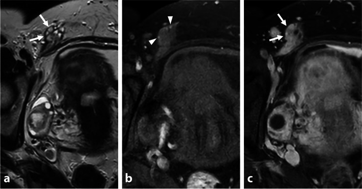
Endometriosis is a common crippling disease in women of reproductive age. Magnetic resonance imaging (MRI) is considered the cornerstone radiological technique for both the diagnosis and management of endometriosis. While its sensitivity, especially in deep infiltrating endometriosis, is superior to that of ultrasonography, many sources of false-positive results exist, leading to a lack of specificity. Hypointense lesions or pseudo-lesions on T2-weighted images include anatomical variants, fibrous connective tissues, benign and malignant tumors, feces, surgical materials, and post treatment scars which may mimic deep pelvic infiltrating endometriosis. False positives can have a major impact on patient management, from diagnosis to medical or surgical treatment. This educational review aims to help the radiologist acknowledge MRI criteria, pitfalls, and the differential diagnosis of deep pelvic infiltrating endometriosis to reduce false-positive results. Critical relevance statement MRI in deep infiltrating endometriosis has a 23% false-positive rate, leading to misdiagnosis. T2-hypointense lesions primarily result from anatomical variations, fibrous connective tissue, benign and malignant tumors, feces, surgical material, and post-treatment scars. Key points • MRI in DIE has a 23% false-positive rate, leading to potential misdiagnosis.• Anatomical variations, fibrous connective tissues, neoplasms, and surgical alterations are the main sources of T2-hypointense mimickers.• Multisequence interpretation, morphologic assessment, and precise anatomic localization are crucial to prevent overdiagnosis.• Gadolinium injection is beneficial for assessing endometriosis differential diagnosis only in specific conditions.

Keywords: Deep infiltrating endometriosis; Endometriosis; Genital diseases; Magnetic resonance imaging; Pelvic inflammatory disease.


Conflict of interest statement

Pascal Rousset reported consultant fees from Ziwig and EDAP TMS France and reported receiving lecture fees from Bracco and compensation for serving on the board from Guerbet.

Stéphanie Nougaret is funded by the European Research Grant (ERC starting grant) and Integrated Cancer Research Grant (SIRIC).

Isabelle Thomassin-Naggara reported receiving lecture fees from General Electric, Siemens, Canon, and GSK; lecture fees and compensation for serving on the board from Guerbet; compensation for serving on the board from Bayer; lecture fees from Incepto, ICAD, Fujifilm, and Hologic; and lecture fees and compensation for serving on the board from Bracco.

Figures

Fig. 1
Fig. 1
Left utero-sacral ligament (USL) varicosity in a 46-year-old woman with chronic catamenial pelvic pain. a Axial, (b) sagittal, and (c) coronal T2-W MR images show a thickened and pseudonodular left USL (arrows) due to tubular and serpiginous T2-hyperintense veinous structures. No pelvic endometriosis was found at surgery; a pelvic venous congestion syndrome was then suggested
Fig. 2
Fig. 2
Thickening of the utero-sacral ligaments (USLs) in a 44-year-old woman with history of PID. Acute episode in 2020:a Axial T2-W MR image shows irregular and pseudo-nodular thickening of bilateral USLs (arrows). b Axial T1-W fat-suppressed contrast-enhanced MR image shows pyosalpinx (star) with thick-walled fallopian tube and surrounding fat stranding. One year follow-up in 2021:c Axial and (d) sagittal T2-W MR images show persistence of pseudonodular thickening of the utero-sacral ligaments (arrows) and the torus, without hemorrhagic foci (not shown)
Fig. 3
Fig. 3
Right round ligament varicosity in the inguinal canal in a 33-year-old woman. a Axial T2-W MR images show pseudocystic changes of the right round ligament in its inguinal course (arrows). Thickening appears regular, without any fluid around the ligament. b Axial fat-suppressed T1-W MR image reveals a few T1-hyperintense foci (arrowheads) within the right round ligament, due to an “entry slice phenomenon artifact”. c Axial T1-weighted fat-suppressed contrast-enhanced MR image shows homogeneous enhancement of tubular veins around the right round ligament (arrows)
Fig. 4
Fig. 4
Fibrous remnant of the urachus insertion in two distinct women of reproductive age. a Sagittal and (b) coronal T2-W MR images show a pseudonodular or triangular T2-hypointense medial structure of the urinary bladder apex at the urachus insertion (arrows). Note the absence of abnormality of the urinary bladder muscular layer (arrowheads), or hemorrhagic foci on T1-WI (no shown)
Fig. 5
Fig. 5
Uterine contraction in a 19-year-old woman with chronic catamenial pelvic pain. a Sagittal T2-W MR image shows a T2-hypointense focal thickening (arrow) of the outer myometrium on the back wall of the uterus. b Note the absence of abnormality of the myometrium and its complete resolution on additional T2-W MR sequences repeated at the end of the exam
Fig. 6
Fig. 6
Anterior adhesions in the vesicouterine pouch after C-section in a 40-year-old woman with medical history of pelvic endometriosis. a Sagittal and (b) axial T2-W MR images show a T2-hypointense fibrous thickening of the vesicouterine pouch, with severe adhesions of the uterine body (arrows). c Axial T1-W fat-suppressed MR image shows no hemorrhagic foci in the anterior subperitoneal space (arrow). Endometriosis surgical management has been decided. During surgical procedure, pelvic anterior symphysis was proven with no obvious endometriosis lesion (confirmed with negative biopsies at pathology)
Fig. 7
Fig. 7
Sporadic pelvic wall desmoid tumor in a 30-year-old woman with history of cesarean section. a Axial T2-W MR image shows an infiltrative mass of the right rectus muscle with heterogeneous T2 signal intensity varying from low (thin arrows) to intermediate (thick arrow) signal intensity areas. Anterior focal adhesions due to previous cesarean section are seen (arrowheads). b Axial diffusion-weighted MR image shows high signal intensity within the mass consistent with increased cellularity (arrows). c Axial T1-W fat-suppressed contrast-enhanced MR image shows heterogeneous enhancement (arrows)
Fig. 8
Fig. 8
Rectal adenocarcinoma in a 33-year-old patient with chronic pelvic pain and rectal disorder with rectal bleeding, addressed for suspicion of endometriosis. a Sagittal, (b) coronal, and (c) axial T2-W MR images show a T2-hypointense focal wall thickening of the high rectum (thick arrows) with a T2-hypointense tumoral infiltration of the mesorectum (thin arrows). Note the absence of retrocervical deep infiltrating endometriosis. d Axial T1-W fat-suppressed contrast-enhanced MR image shows a moderately enhanced tumor (arrows)
Fig. 9
Fig. 9
Actinomycosis in a 36-year-old woman with chronic pelvic pain, rectal disorder. and dyspareunia. a Axial and (b) sagittal T2-W MR images show right subperitoneal infiltration with intermediate signal intensity, centered on the right utero-sacral ligament (thin arrows) and the sacro-recto-genital septum up to the presacral space (star) from the first sacral vertebra to the sacrococcygeal junction. c Axial T2-W MR image shows perirectal soft-tissue infiltration (arrows) with intermediate T2-signal intensity semicircumferential thickening of the upper and posterior rectum (arrowheads). d Axial T1-W fat-suppressed contrast-enhanced subtracted MR image shows avid enhancement of the surrounding inflammatory infiltration with small abscesses in the presacral space (arrows). Past medical history of intrauterine device recent removal was found
Fig. 10
Fig. 10
Pelvic alveolar echinococcosis in a 28-year-old woman with chronic pelvic pain, bladder disorder, and dyspareunia. a Sagittal and (b) axial T2-W MR images show an infiltrative T2-hypointense external myometrial infiltrative mass (thick arrows) with a few microcysts (arrowheads), and a contiguous infiltration of the bladder dome (thin arrows). c Axial T1-W fat-suppressed MR image shows several microcysts (arrowheads) without hemorrhagic foci in the extrinsic infiltrative uterine mass. d Axial T1-W fat-suppressed contrast-enhanced subtracted MR image shows central necrosis (arrows) and irregular margin with a peripheral enhancement. History of liver alveolar echinococcosis infection in childhood was then found
Fig. 11
Fig. 11
Normal sigmoid colon filled with feces in a 35-year-old woman with chronic pelvic pain. No medical history. a Axial and (b) coronal T2-W MR images show a T2-hypointense “pseudo”-thickening of the posterior sigmoid colon wall (arrows). Note the absence of retrocervical deep infiltrating endometriosis (arrowheads). c Axial T1-W fat-suppressed MR image shows the absence of hyperintense foci nor abnormality of the sigmoid colon wall (arrow), with a more or less fecal content. Laparoscopy showed a normal recto-sigmoid colon
Fig. 12
Fig. 12
Unilateral right-sided vesicoureteral reflux surgical bulking agent (Macroplastique®—polydimethylsiloxane injection) in a 26-year-old woman. a Sagittal and (b) axial T2-W MR images show a T2-hypointense unilateral right-sided vesicoureteral reflux implant (arrows) at the ureterovesical junction. c Axial T1-W fat-suppressed MR image shows an ovoid geometrical shape in T1-isosignal intensity (arrow)

References

    1. Taylor HS, Kotlyar AM, Flores VA. Endometriosis is a chronic systemic disease: clinical challenges and novel innovations. Lancet. 2021;397:839–852. doi: 10.1016/S0140-6736(21)00389-5. - DOI - PubMed
    1. International working group of AAGL, ESGE, ESHRE and WES. Tomassetti C, Johnson NP, et al. An international terminology for endometriosis, 2021. J Minim Invasive Gynecol. 2021;28:1849–1859. doi: 10.1016/j.jmig.2021.08.032. - DOI - PubMed
    1. Nisenblat V, Bossuyt PMM, Farquhar C, et al. Imaging modalities for the non-invasive diagnosis of endometriosis. Cochrane Database Syst Rev. 2016;2:CD009591. doi: 10.1002/14651858.CD009591.pub2. - DOI - PMC - PubMed
    1. (2012) Endometriosis and infertility: a committee opinion. Fertil Steril 98:591–598. 10.1016/j.fertnstert.2012.05.031 - PubMed
    1. Chapron C, Marcellin L, Borghese B, Santulli P. Rethinking mechanisms, diagnosis and management of endometriosis. Nat Rev Endocrinol. 2019;15:666–682. doi: 10.1038/s41574-019-0245-z. - DOI - PubMed

LinkOut - more resources