

Comparison of three-dimensional imaging of the nose using three different 3D-photography systems: an observational study
- PMID: 38267982
- PMCID: PMC10807178
- DOI: 10.1186/s13005-024-00406-4
Comparison of three-dimensional imaging of the nose using three different 3D-photography systems: an observational study
Abstract
Background: New 3D technologies for superficial soft tissue changes, especially in plastic and reconstructive surgical procedures, can improve the planning and documentation of facial surgeries. The purpose of this study was to compare and determine the applicability and feasibility of three different 3D-photography systems in clinical practice imaging the nose.
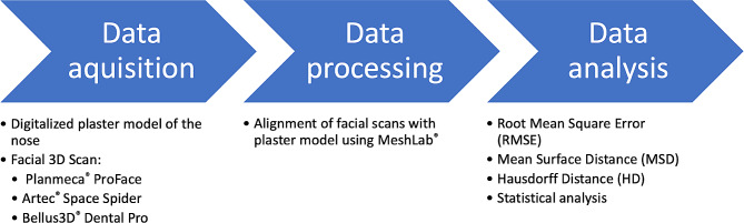
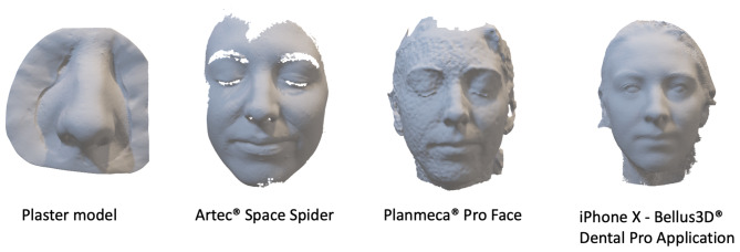
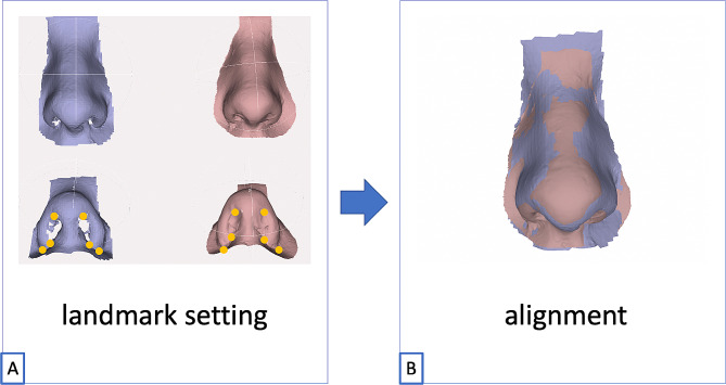
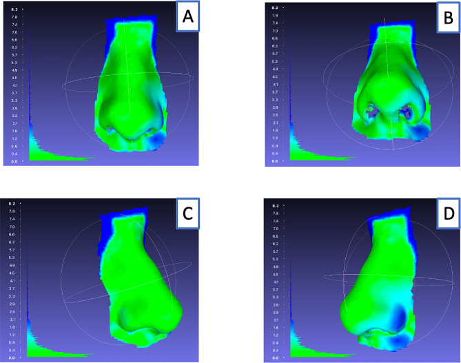
Methods: A total of 16 healthy non-operated noses were included in this prospective study. A plaster model of each nose was produced, digitized, and converted to a .stl mesh (= ground truth model). Three-dimensional images of each nose were then taken using Artec Space Spider (gold standard), Planmeca ProFace®, and the Bellus3D Dental Pro application. All resulting .stl files were aligned to the ground truth model using MeshLab software, and the root mean square error (RMSE), mean surface distance (MSD), and Hausdorff distance (HD) were calculated.
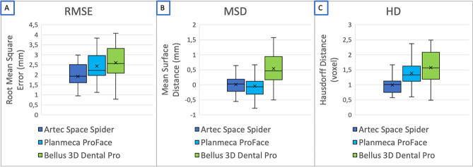
Results: The Artec Space Spider 3D-photography system showed significantly better results compared to the two other systems in regard to RMSE, MSD, and HD (each p < 0.001). There was no significant difference between Planmeca ProFace® and Bellus3D Dental Pro in terms of RMSE, MSD, and HD. Overall, all three camera systems showed a clinically acceptable deviation to the reference model (range: -1.23-1.57 mm).
Conclusions: The three evaluated 3D-photography systems were suitable for nose imaging in the clinical routine. While Artec Space Spider showed the highest accuracy, the Bellus3D Dental Pro app may be the most feasible option for everyday clinical use due to its portability, ease of use, and low cost. This study presents three different systems, allowing readers to extrapolate to other systems when planning to introduce 3D photography in the clinical routine.
Keywords: 3D photography; 3D technologies, Face scan, TrueDepth; Rhinoplasty.
© 2024. The Author(s).
Conflict of interest statement
The authors declare no competing interests. The authors declare that the research was conducted in the absence of any commercial or financial relationships that could be construed as a potential conflict of interest.
Figures
References
-
- Koban KC, Perko P, Etzel L, Li Z, Schenck TL, Giunta RE. Validation of two handheld devices against a non-portable three-dimensional surface scanner and assessment of potential use for intraoperative facial imaging. J Plast Reconstr Aesthetic Surg. 2020;73(1):141–8. doi: 10.1016/j.bjps.2019.07.008. - DOI - PubMed
-
- Papadopoulos MA, Christou PK, Christou PK, Athanasiou AE, Boettcher P, Zeilhofer HF, Three-dimensional craniofacial reconstruction imaging. Oral Surgery, Medicine O et al. Oral Pathology, Oral Radiology, and Endodontology. 2002;93(4):382–93. - PubMed
Publication types
MeSH terms
LinkOut - more resources
Full Text Sources
Medical
