. 2024 Jan 25;21(1):2.
doi: 10.1186/s12983-024-00521-7.

New insights into mesoderm and endoderm development, and the nature of the onychophoran blastopore

Affiliations

New insights into mesoderm and endoderm development, and the nature of the onychophoran blastopore

Ralf Janssen et al. Front Zool. .

Abstract

Background: Early during onychophoran development and prior to the formation of the germ band, a posterior tissue thickening forms the posterior pit. Anterior to this thickening forms a groove, the embryonic slit, that marks the anterior-posterior orientation of the developing embryo. This slit is by some authors considered the blastopore, and thus the origin of the endoderm, while others argue that the posterior pit represents the blastopore. This controversy is of evolutionary significance because if the slit represents the blastopore, then this would support the amphistomy hypothesis that suggests that a slit-like blastopore in the bilaterian ancestor evolved into protostomy and deuterostomy.

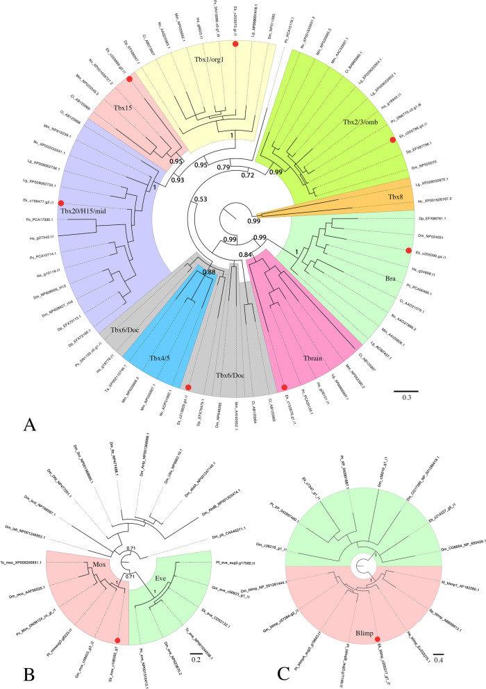
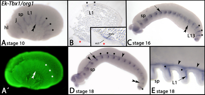
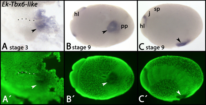
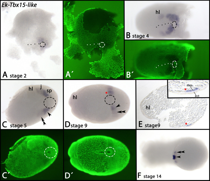
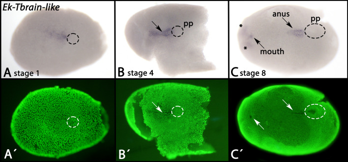
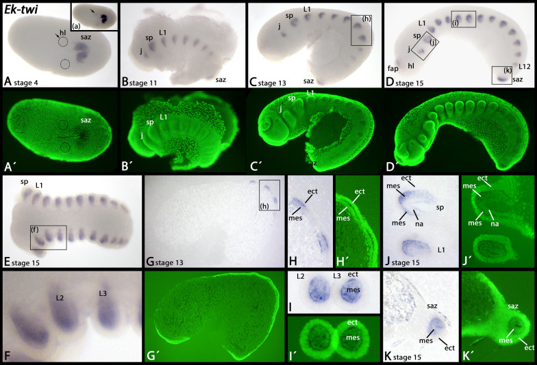
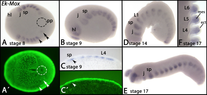
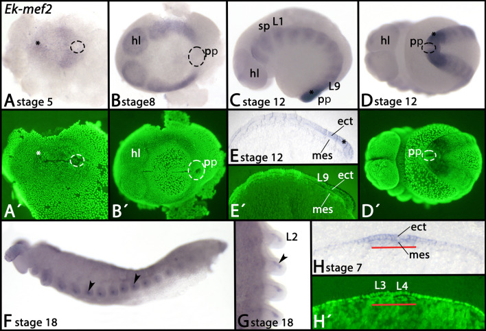
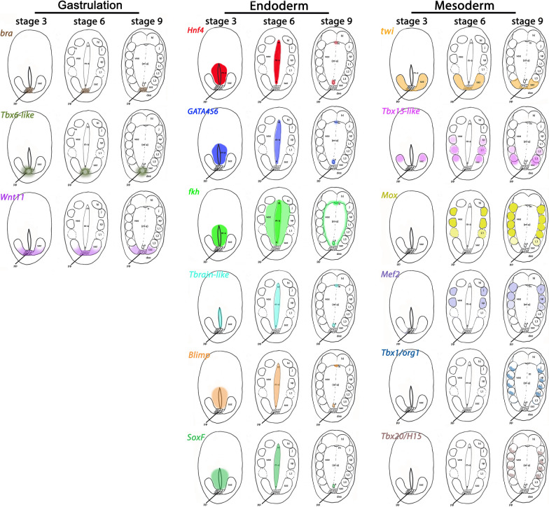
Results: In this paper, we summarize our current knowledge about endoderm and mesoderm development in onychophorans and provide additional data on early endoderm- and mesoderm-determining marker genes such as Blimp, Mox, and the T-box genes.

Conclusion: We come to the conclusion that the endoderm of onychophorans forms prior to the development of the embryonic slit, and thus that the slit is not the primary origin of the endoderm. It is thus unlikely that the embryonic slit represents the blastopore. We suggest instead that the posterior pit indeed represents the lips of the blastopore, and that the embryonic slit (and surrounding tissue) represents a morphologically superficial archenteron-like structure. We conclude further that both endoderm and mesoderm development are under control of conserved gene regulatory networks, and that many of the features found in arthropods including the model Drosophila melanogaster are likely derived.

Keywords: Archenteron; Blastopore; Blimp; Mox; Onychophora; T-box transcription factor; Twist; mef2.


Conflict of interest statement

The authors declare that they have no competing interests.

Figures

Fig. 1
Fig. 1
Phylogenetic analyses Phylogenetic trees of T-box genes A, Mox genes B and Blimp genes C. Bayesian analyses using MrBayes [39] applying 0.75 million cycles (T-box Tree), 1 million cycles (Mox tree), and 0.3 million cycles (Blimp tree) for the Metropolis-Coupled Markov Chain Monte Carlo (MCMCMC). The scale bars represent the amino acid substitutions rate per site. Orthologs of Mox proteins are compared with Eve genes and Hox genes B. Orthologs of Blimp genes are compared with the most closely related genes of these species, many of them representing previously uncharacterized genes C. Support values represent posterior probabilities. Species abbreviations: Ci; Ciona intestinalis (Chordata); Dm, Drosophila melanogaster (Arthropoda: Insecta); Dp, Daphnia pulex (Arthropoda: Branchiopoda); Ek, Euperipatoides kanangrensis (Onychophora); Gm, Glomeris marginata (Arthropoda: Myriapoda); Hs, Halicryptus spinulosus (Priapulida); He, Hydroides elegans (Polychaeta); Lg, Lottia gigantea (Mollusca); Mm, Mus musculus (Vertebrata); Nv, Nematostella vectensis (Cnidaria); Pc, Priapulus caudatus (Priapulida); Parasteatoda tepidariorum (Arthropoda: Chelicerata); Sp, Strongylocentrotus purpuratus (Echinodermata); Ta, Trichoplax adhaerens (Placozoa); Tc, Tribolium castaneum (Arthropoda: Insecta); Xl, Xenopus laevis (Vertebrata). Accession numbers are listed in (Additional file 7: Supplementary File 7)
Fig. 2
Fig. 2
Expression of Tbx1/org1 In all panels, anterior is to the left. Panels A, C and D represent lateral views, dorsal up. Panel B represents a thin section. Panel E represents a dorsal view. Panel A´ represents a SYBR Green staining of the embryo shown in panel A. The double arrowheads in panels A and C point to ventral expression. The asterisks in panels A-D mark segmental expression in newly formed segments. The red asterisk in B marks the enlarged region shown in the inlay in B. The arrow in panels C and E point to expression in the limbs. Arrowheads in panels D and E point to dorsal expression. Filled circles in panel D and E mark patches of stronger expression dorsal to each appendage. Developmental stages are indicated (staging system after [9], their supplementary data). Abbreviations: ect, ectoderm; hl, head lobe; j, jaw; L, leg; mes, mesoderm; sp, slime papilla
Fig. 3
Fig. 3
Expression of Tbx6-like In all panels, anterior is to the left. Panels A and B represent ventral views. Panel C represents a lateral view, dorsal up. Panels A´-C´ represent SYBR Green staining of the embryos shown in panels A-C. The dotted line in panel A marks the embryonic slit. The arrowheads in panels A-C point to expression in the ectoderm of the posterior pit. Developmental stages are indicated (staging system after [9], their supplementary data). Abbreviations: hl, head lobe; j, jaw; pp, posterior pit; slime papilla
Fig. 4
Fig. 4
Expression of Tbx15-like In all panels, anterior is to the left. Panels A–D and F represent ventral views. Panel E represents a thin section, lateral view, dorsal up. Panels A´–D´ represent SYBR Green staining of the embryos shown in panels A–D. The dashed circles in panels A–D mark the posterior pit region. The dashed line in panels A and B mark the embryonic slit. The red asterisk in panels D and E mark the position in a whole mount (D) and a thin section E. Arrowheads and double arrowheads in panels C, D and F mark expression in the last-formed posterior somites. Developmental stages are indicated (staging system after [9], their supplementary data). Abbreviations: ect, ectoderm; hl, head lobe; j, jaw; mes, mesoderm; sp, slime papilla
Fig. 5
Fig. 5
Expression of Tbx20/H15 In all panels, anterior is to the left, lateral views, dorsal up. Panels A´ and D´ represent SYBR Green staining of the embryos shown in panels A and D. Asterisks in panels A and B mark segmental expression. Arrows in panels B–D point to mesodermal expression in the developing appendages. Panel D represents a thin section. The arrowhead in panel E marks dorsal expression in the developing heart. Developmental stages are indicated (staging system after [9], their supplementary data). Abbreviations: e, eye; hl, head lobe; j, jaw; L, leg; sp, slime papilla
Fig. 6
Fig. 6
Expression of Tbrain-like In all panels, anterior is to the left, ventral views. Panels A´–C´ represent SYBR Green staining of the embryos shown in panels A–C. Arrows point to expression in the embryonic slit. Dashed circles mark the position of the posterior pit. Asterisks in panel C mark expression in the the anterior tissue of the developing embryo that could be associated with the developing brain. Developmental stages are indicated (staging system after [9], their supplementary data). Abbreviations: pp, posterior pit
Fig. 7
Fig. 7
Expression of twi In all panels, anterior is to the left, except panel J (anterior up). Panels A represents a ventral view, all other panels represent lateral views (dorsal up). Section (a) (inlay in A) shows the same embryo as in A with increased contrast to better visualize the weak most anterior expression in the somite corresponding to the head lobes (hl) (arrow). The box in panel C marks the same area as shown in panels G and H (thin sections). Panel H shows a magnification of the boxed area in panel G. Boxes in panel D show same areas as in panels I-K (thin sections). Panels A´–D´ and G´–K represent SYBR Green staining of the embryos shown in corresponding panels. Developmental stages are indicated (staging system after [9], their supplementary data). Abbreviations: ect, ectoderm; fap, frontal appendage; hl, head lobe; j, jaw; L, leg; mes, mesoderm; na, nephridial anlagen; saz, segment addition zone; sp, slime papilla
Fig. 8
Fig. 8
Expression of Mox In all panels, anterior is to the left, except panel F (dorsal view, anterior up). Panel A represents a ventral view. Panels B–E represent lateral views, dorsal up Panels A´ and C´ represent SYBR Green staining of the embryos shown in panels A and C. The dashed circle in panel A marks the region of the posterior pit. The arrow and arrowhead in panel A point to expression in the last-formed posterior somites. The arrowhead in panel C (thin section) points to expression in the mesoderm. Developmental stages are indicated (staging system after [9], their supplementary data). Abbreviations: ect, ectoderm; hl, head lobe; j, jaw; L, leg; mes, mesoderm; pp, posterior pit; sp, slime papilla
Fig. 9
Fig. 9
Expression of mef2 In all panels anterior is to the left, except panel G (anterior up). Panels A, B, D and G represent ventral views. Panels C, E (thin section), F and H (thin section) represent lateral views, dorsal up. Panels A´, B´, D´, E´ and H´ represent SYBR Green staining of the embryos shown in corresponding panels. Dashed circles in panels A, B and D mark the posterior pit region. The asterisk in panel A marks enhanced expression anterior in the embryonic slit. The asterisks in panels C–E mark ectodermal expression overlying the last-formed posterior somite(s). Arrowheads in panels F and G point to expression in the mesoderm of developing appendages. The red bar in panels H/H´ mark the area of mesodermal expression. Developmental stages are indicated (staging system after [9], their supplementary data). Abbreviations: ect, ectoderm; hl, head lobe; L, leg; mes, mesoderm; pp, posterior pit; sp, slime papilla
Fig. 10
Fig. 10
Expression of nau In panels A and B anterior is to the left; in panel C anterior is up. Panels A and B represent lateral views, panel C shows as ventral view. The arrowheads point to expression in the mesoderm of developing appendages. Developmental stages are indicated (staging system after [9], their supplementary data). Abbreviations: j, jaw; hl, head lobe; L, leg; sp, slime papilla
Fig. 11
Fig. 11
Expression of Blimp In all panels, anterior is to the left and ventral views. Panels A´–D´ represent SYBR Green staining of the embryos shown in panels A–D. Dashed lines mark the position of the posterior pit region. Arrowheads point to expression in the embryonic slit. The asterisk in panel D marks the mouth that does not express Blimp in later developmental stages. Developmental stages are indicated (staging system after [9], their supplementary data). Abbreviations: hl, head lobe
Fig. 12
Fig. 12
Expression of SoxF In all panels, anterior is to the left, ventral views. Panels A´–C´ represent SYBR Green staining of the embryos shown in panels A–C. Dashed circles mark the posterior pit region. Arrows in panel A point to scattered cells that express SoxF. Arrowheads point to expression in the embryonic slit. In all panels, the posterior pit is encircled. Developmental stages are indicated (staging system after [9], their supplementary data). Abbreviations: hl, head lobe
Fig. 13
Fig. 13
Schematic overview, expression of endoderm- and mesoderm-patterning genes The early expression of marker genes is shown in gene-specific colours. In the schematic drawings of embryos, anterior is pointing upwards, ventral views. Abbreviations: a, anus; dee, dorsal extraembryonic tissue; hl, somite of the head lobe segment; j, somite of the jaw-bearing segment; L, somite of a leg-bearing segment; m, mouth; m-a, mouth-anus furrow (the embryonic slit); pp, posterior pit (blastopore); saz, segment addition zone; sp, somite of the slime papilla-bearing segment; vee, ventral extraembryonic tissue

Similar articles

Cited by

References

    1. Wu R, Pisani D, Donoghue PCJ. The unbearable uncertainty of panarthropod relationships. Biol Lett. 2023;19(1):20220497. doi: 10.1098/rsbl.2022.0497. - DOI - PMC - PubMed
    1. Nájera GS, Weijer CJ. The evolution of gastrulation morphologies. Development. 2023;150(7):dev200885. doi: 10.1242/dev.200885. - DOI - PMC - PubMed
    1. Budd GE, Jensen S. A critical reappraisal of the fossil record of the bilaterian phyla. Biol Rev. 2000;70:253–295. doi: 10.1111/j.1469-185X.1999.tb00046.x. - DOI - PubMed
    1. Technau U, Scholz CB. Origin and evolution of endoderm and mesoderm. Int J Dev Biol. 2003;47(7–8):531–539. - PubMed
    1. Manton SM. Studies on the Onychophora VII. The early embryonic stages of Peripatopsis, and some general considerations concerning the morphology and phylogeny of the arthropoda. Philos Trans R Soc B-Biol Sci. 1949;233:483–580.

LinkOut - more resources