. 2024 Jan 24;22(1):99.
doi: 10.1186/s12967-024-04897-2.

Fbxw7 suppresses carcinogenesis and stemness in triple-negative breast cancer through CHD4 degradation and Wnt/β-catenin pathway inhibition

Affiliations

Fbxw7 suppresses carcinogenesis and stemness in triple-negative breast cancer through CHD4 degradation and Wnt/β-catenin pathway inhibition

Guodong Xiao et al. J Transl Med. .

Abstract

Background: Cancer stem cells (CSCs) are a small population of cells in tumor tissues that can drive tumor initiation and promote tumor progression. A small number of previous studies indirectly mentioned the role of F-box and WD repeat domain-containing 7 (FBXW7) as a tumor suppressor in Triple-negative breast cancer (TNBC). However, few studies have focused on the function of FBXW7 in cancer stemness in TNBC and the related mechanism.

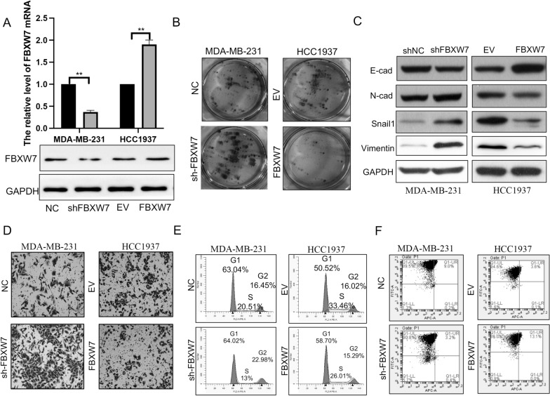
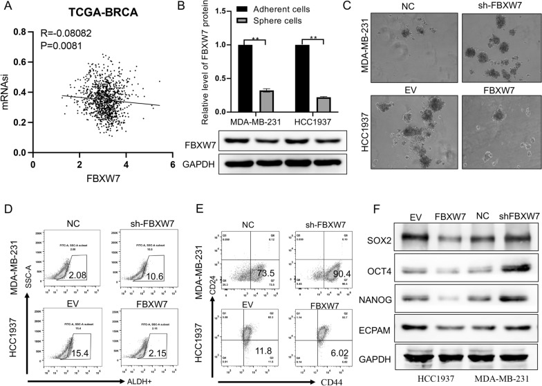
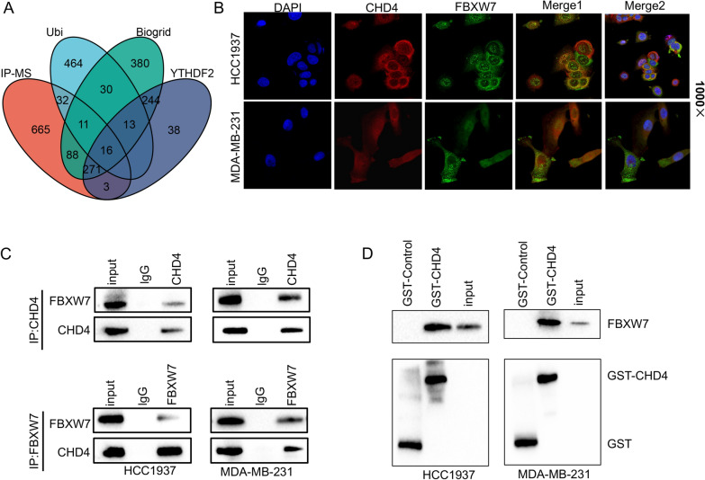
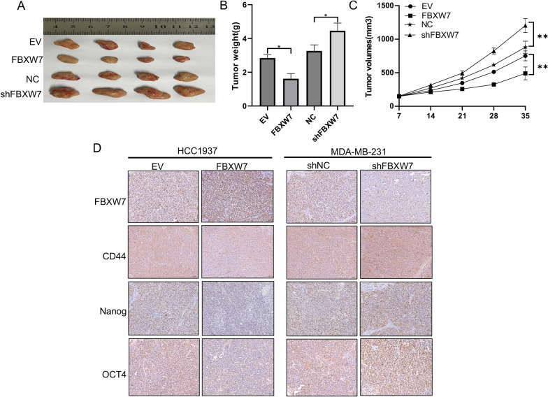
Methods: We detected FBXW7 by immunohistochemistry (IHC) in 80 TNBC patients. FBXW7 knockdown and overexpression in MD-MBA-231 and HCC1937 cell models were constructed. The effect of FBXW7 on malignant phenotype and stemness was assessed by colony assays, flow cytometry, transwell assays, western blot, and sphere formation assays. Immunoprecipitation-Mass Spectrometry (IP-MS) and ubiquitination experiments were used to find and verify potential downstream substrate proteins of FBXW7. Animal experiments were constructed to examine the effect of FBXW7 on tumorigenic potential and cancer stemness of TNBC cells in vivo.

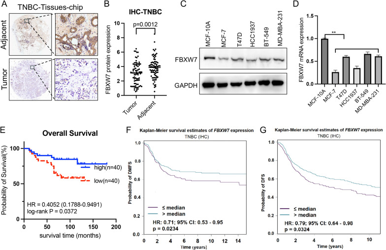
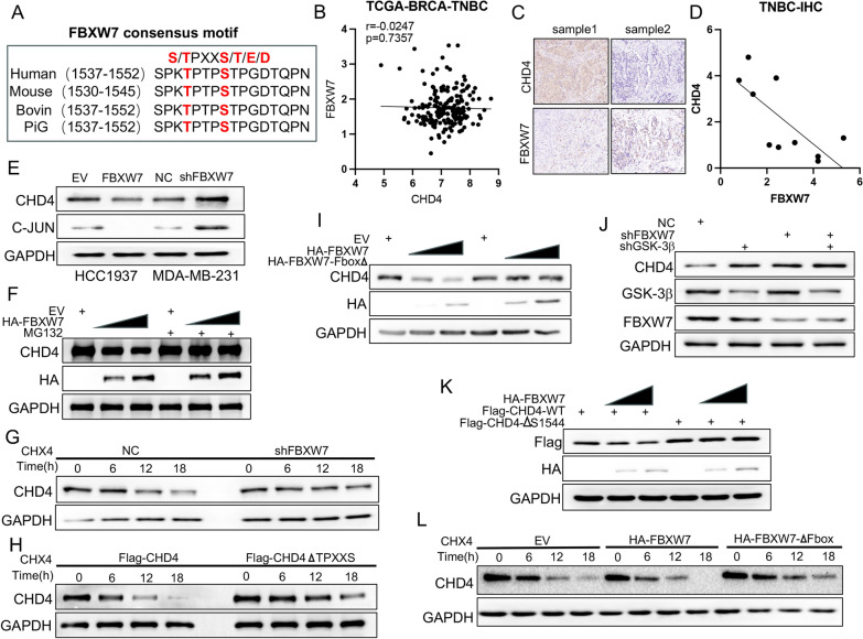
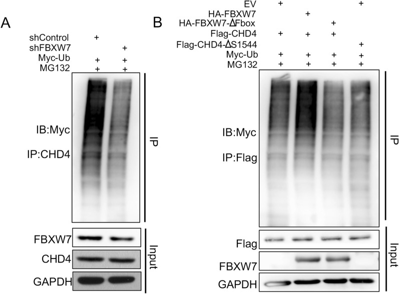
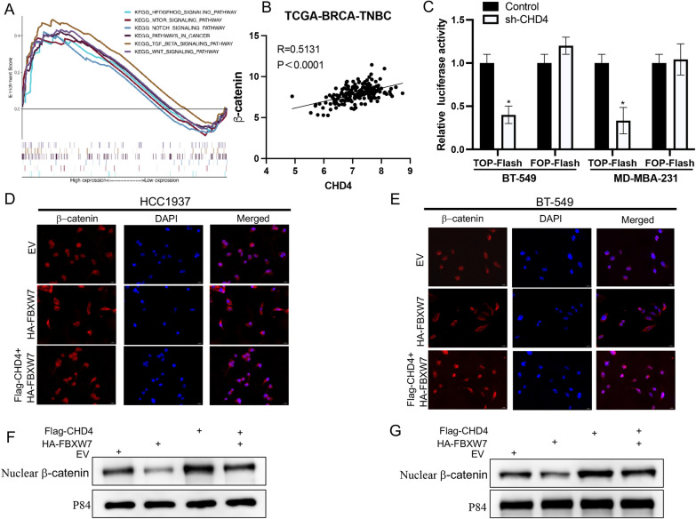
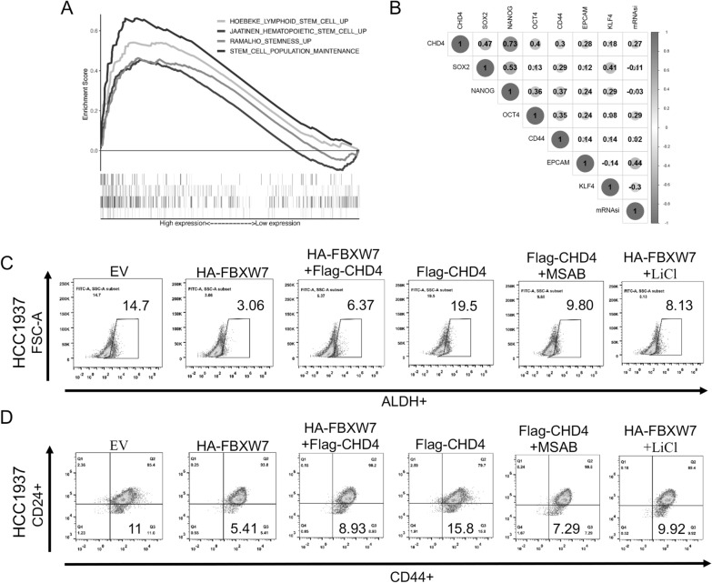
Results: The results showed that FBXW7 was expressed at low levels in TNBC tissues and positively correlated with prognosis of TNBC patients. In vitro, FBXW7 significantly inhibited colony formation, cell cycle progression, cell migration, EMT process, cancer stemness and promotes apoptosis. Further experiments confirmed that chromodomain-helicase-DNA-binding protein 4 (CHD4) is a novel downstream target of FBXW7 and is downregulated by FBXW7 via proteasomal degradation. Moreover, CHD4 could promote the nuclear translocation of β-catenin and reverse the inhibitory effect of FBXW7 on β-catenin, and ultimately activate the Wnt/β-catenin pathway. Rescue experiments confirmed that the FBXW7-CHD4-Wnt/β-catenin axis was involved in regulating the maintenance of CSC in TNBC cells. In animal experiments, FBXW7 reduced CSC marker expression and suppressed TNBC cell tumorigenesis in vivo.

Conclusions: Taken together, these results highlight that FBXW7 degrades CHD4 protein through ubiquitination, thereby blocking the activation of the Wnt/β-catenin pathway to inhibit the stemness of TNBC cells. Thus, targeting FBXW7 may be a promising strategy for therapeutic intervention against TNBC.

Keywords: CHD4; Fbxw7; Stemness; Triple-negative breast cancer; Wnt/β-catenin.


Conflict of interest statement

The authors have declared no competing interests.

Figures

Fig. 1
Fig. 1
Low expression of FBXW7 in TNBC patients is associated with a poor prognosis. A Representative images of immunohistochemical staining of FBXW7 in TNBC tissues and corresponding adjacent noncancerous tissues using TMA tissue sections (n = 80 TNBC). B Quantitative values for IHC staining of FBXW7 between tumor tissues and adjacent nontumor normal tissues are presented in the histogram. Statistical analysis was performed by two-tailed paired Student’s t test (p = 0.0012). C, D Western blot and qRT‒PCR analysis of FBXW7 expression in mammary epithelial cell lines (MCF-10A), two non-TNBC cell lines (MCF-7 and T47-D) and three TNBC cell lines (HCC1937, BT549 and MD-MBA-231). E Kaplan–Meier survival curves for OS of TNBC patients in the high FBXW7 and low FBXW7 groups. F, G Kaplan‒Meier survival curves of DMFS and DFS based on FBXW7 expression using the bc-GenExMiner online tool (http://bcgenex.ico.unicancer.fr/BC-GEM/GEM-Accueil.php?js=1); HR values are shown
Fig. 2
Fig. 2
FBXW7 suppresses cell proliferation, cell cycle progression, apoptosis, and EMT in TNBC cells. A Western blot and qRT‒PCR analysis of FBXW7 expression following FBXW7 knockdown in MD-MBA-231 cells and FBXW7 overexpression in HCC1937 cells. ***p < 0.0001. B Colony formation assays were performed to measure the proliferation ability of the transfected TNBC cells. C The levels of EMT marker proteins in the transfected TNBC cells were detected using Western blot analysis. D Transwell assays were performed to determine the migration abilities after FBXW7 knockdown and overexpression in MD-MBA-231 and HCC1937 cells, respectively. E, F FACS assays were performed on the transfected TNBC cells to analyze the cell cycle and apoptosis
Fig. 3
Fig. 3
FBXW7 overexpression inhibits the stem cell-like properties of TNBC cells. A Pearson’s correlation coefficient analysis of the correlation analysis between FBXW7 and mRNAsi in TCGA-BRCA datasets (r = − 0.08082, p = 0.0081). B FBXW7 expression was detected in spheres and their adherent TNBC cells by Western blotting. C Representative images and quantitative analysis of spheres. Scale bar: 100 mm. D ALDEFLUOR assay of the percentage of ALDH-positive cells in different treatment groups. E FACS profiles and quantification of the CD44high/CD24low subpopulation. F Western blot analysis of the protein levels of stem cell markers (SOX2, OCT4, NANOG and EPCAM) in different cell treatment groups
Fig. 4
Fig. 4
CHD4 is a binding partner of FBXW7. A Venn diagram showing the number of overlapping genes between the four datasets as indicated. B Intracellular localization analysis of FBXW7 and CHD4 by immunofluorescence assay. The intracellular localization of CHD4 (red) and FBXW7 (green) is shown. Nuclei (blue) were stained with 4′,6-diamidino-2-phenylindole (DAPI). C Co-IP assays and Western blotting were conducted to detect the relationship between CHD4 and FBXW7. D A GST pull-down assay and Western blotting were performed to determine the interaction between FBXW7 and CHD4
Fig. 5
Fig. 5
CHD4 protein stability is regulated by FBXW7-mediated ubiquitination. A Sequence alignment of the phosphodegron sequences recognized by FBXW7 in CHD4. Conserved degron sequences are shown in red. B Scatter diagram showing the correlation between the RNA expression levels of CHD4 and FBW7 in TNBC patients based on TCGA-BRCA-TNBC datasets. P values are indicated. C Representative IHC images of FBXW7 and CHD4 staining. The scale bar indicates 50 μm. D The correlations between the protein expression of CHD4 and FBXW7 in TNBC tissues. E Western blot analysis of the regulation of CHD4 and JUN protein expression by FBXW7 in two transfected TNBC cell lines. F Western blot analysis of CHD4 protein and HA-tag protein in HCC1937 cells with different treatments. which was indicated in the figure labels. G MDA-MB-231 cells were transfected with shNC or shFBXW7 plasmid and evaluated for CHD4 protein half-life by CHX chase assay. H MDA-MB-231 cells were transfected with Flag-CHD4 or Flag-CHD4 ∆TPXXS plasmids and evaluated for CHD4 protein half-life by CHX chase assay. I Western blot analysis of CHD4 protein and HA-tag protein in HCC1937 cells transfected with HA-FBXW7 or HA-FBXW7 ∆Fbox plasmid. J MDA-MB-231 cells were infected with the indicated constructs for 48 h. Cells were subjected to Western blot analysis. K Western blot analysis of Flag-tag protein and HA-tag protein in HCC1937 cells transfected with indicated plasmid. L MDA-MB-231 cells were transfected with empty vector, HA-FBXW7 or HA-FBXW7∆Fbox plasmid and evaluated for CHD4 protein half-life by CHX chase assay. GAPDH served as an internal reference in all of the above Western blot analyses
Fig. 6
Fig. 6
FBW7 serves as an E3 ubiquitin ligase for the CHD4 protein. A, B Western blot analysis of whole cell lysates (WCLs) and immunoprecipitates derived from HEK-293 T cells transfected with the indicated plasmids for 24 h; cells were treated with 20 μM MG132 for an additional 5 h before harvesting. The ubiquitination status of endogenous CHD4 protein or Flag-tag protein was determined by immunoprecipitation (IP). GAPDH was used as an equal loading control
Fig. 7
Fig. 7
The FBXW7/CHD4 axis regulates the Wnt/β-catenin pathway in TNBC cells. A Enrichment plots from GSEA to examine the correlation between CHD4 expression and multiple cancer-related pathways based on TCGA-BRCA-TNBC datasets. B Pearson’s correlation analysis of the relationship between β-catenin expression and CHD4 expression was performed. (r = 0.5131, p < 0.0001). C The TOP/FOP assay was applied to measure the activity of the Wnt signaling pathway in cells transfected with shControl or shCHD4. D, E Immunofluorescence assay examining the nuclear translocation of β-catenin upon treatment with the indicated shRNA. Cell nuclei are stained with DAPI dye (blue). β-Catenin fluorescence intensity was analyzed and quantified by ImageJ (right panel) (200 ×). F, G Western blot analysis was used to examine the β-catenin protein level in the nuclear fraction (nucleus). The quantification of β-catenin protein levels was normalized to that of p84 (nucleus)
Fig. 8
Fig. 8
The FBXW7/CHD4/Wnt/β-catenin axis regulates the stem cell-like properties of TNBC cells. A Enrichment plots from GSEA to examine the correlation between CHD4 expression and multiple stemness-related gene sets based on TCGA-BRCA-TNBC datasets. B Heatmap analysis of the correlation between CHD4 expression and multiple stemness-related genes based on TCGA-BRCA-TNBC datasets. C ALDEFLUOR assay of the percentage of ALDH-positive cells in different treatment groups. D FACS profiles and quantification of the CD44high/CD24low subpopulation in different treatment groups
Fig. 9
Fig. 9
FBXW7 inhibits tumorigenesis in vivo. A Representative images of xenografts in nude mice established via subcutaneous injection of TNBC cells. B, C Growth curve and histogram analysis of the volumes and weights of xenograft tumors. **p < 0.001. D Representative IHC staining images of FBXW7, CD44, Nanog and OCT4 in xenograft tumor tissues

Similar articles

Cited by

References

    1. Dass SA, Tan KL, Selva Rajan R, Mokhtar NF, Mohd Adzmi ER, Wan Abdul Rahman WF, Tengku Din TADA-A, Balakrishnan V. Triple negative breast cancer: A review of present and future diagnostic modalities. Medicina. 2021;57:62. doi: 10.3390/medicina57010062. - DOI - PMC - PubMed
    1. Park S-Y, Choi J-H, Nam J-S. Targeting cancer stem cells in triple-negative breast cancer. Cancers. 2019;11:965. doi: 10.3390/cancers11070965. - DOI - PMC - PubMed
    1. Najafi M, Farhood B, Mortezaee K. Cancer stem cells (CSCs) in cancer progression and therapy. J Cell Physiol. 2019;234:8381–8395. doi: 10.1002/jcp.27740. - DOI - PubMed
    1. He L, Wick N, Germans SK, Peng Y. The role of breast cancer stem cells in chemoresistance and metastasis in triple-negative breast cancer. Cancers. 2021;13:6209. doi: 10.3390/cancers13246209. - DOI - PMC - PubMed
    1. Zhang X, Powell K, Li L. Breast cancer stem cells: biomarkers, identification and isolation methods, regulating mechanisms, cellular origin, and beyond. Cancers. 2020;12:3765. doi: 10.3390/cancers12123765. - DOI - PMC - PubMed

Publication types

MeSH terms

Substances

LinkOut - more resources