. 2023 Nov 4;8(1):102253.
doi: 10.1016/j.rpth.2023.102253. eCollection 2024 Jan.

In vitro characterization of rare anti-αIIbβ3 isoantibodies produced by patients with Glanzmann thrombasthenia that severely block fibrinogen binding and generate procoagulant platelets via complement activation

Affiliations

In vitro characterization of rare anti-αIIbβ3 isoantibodies produced by patients with Glanzmann thrombasthenia that severely block fibrinogen binding and generate procoagulant platelets via complement activation

Christine S M Lee et al. Res Pract Thromb Haemost. .

Abstract

Background: Glanzmann thrombasthenia (GT) is a rare bleeding disorder caused by inherited defects of the platelet αIIbβ3 integrin. Platelet transfusions can be followed by an immune response that can block integrin function by interfering with fibrinogen binding.

Objectives: In this study, we aimed to determine the prevalence of such isoantibodies and better characterize their pathogenic properties.

Methods: Twelve patients with GT were evaluated for anti-αIIbβ3 isoantibodies. Sera from patients with GT with or without anti-αIIbβ3 isoantibodies were then used to study their in vitro effect on platelets from healthy donors. We used several approaches (IgG purification, immunofluorescence staining, and inhibition of signaling pathways) to characterize the pathogenic properties of the anti-αIIbβ3 isoantibodies.

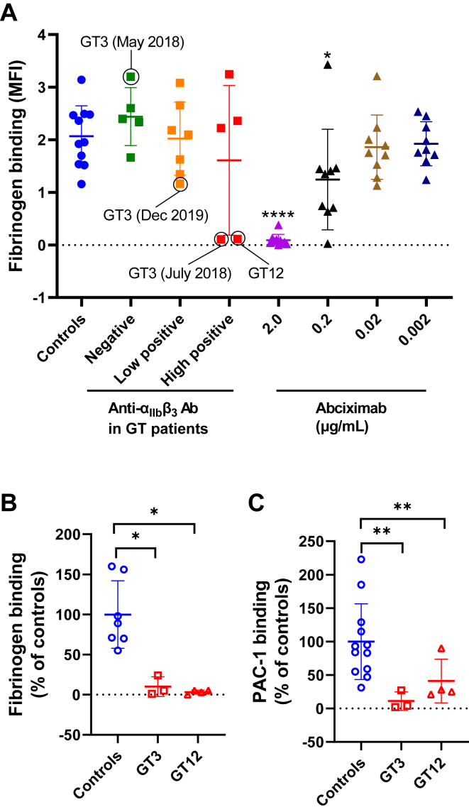
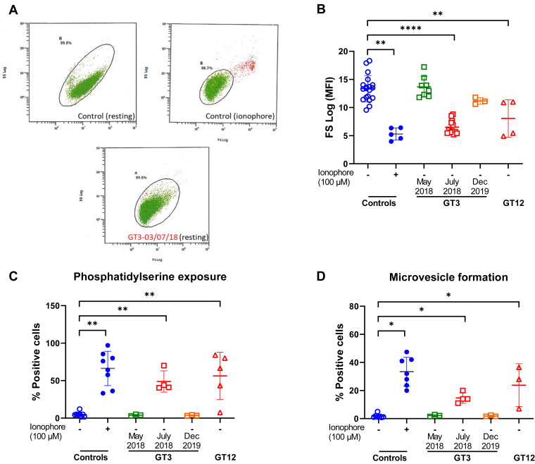
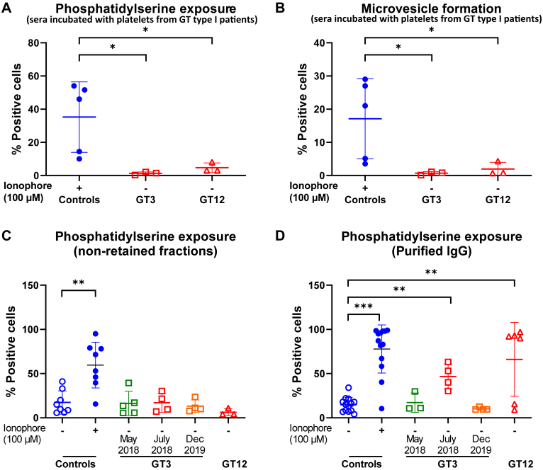
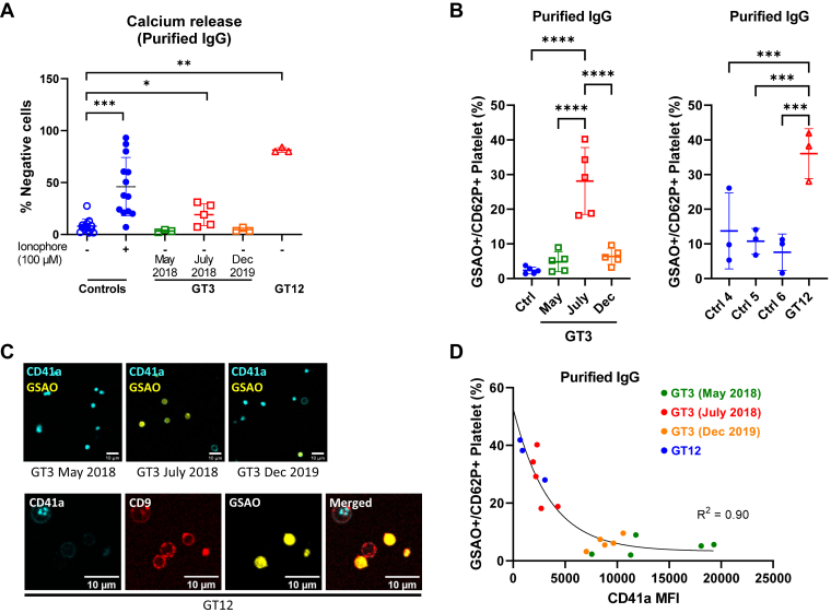
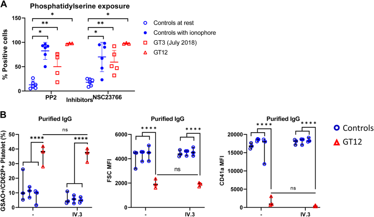
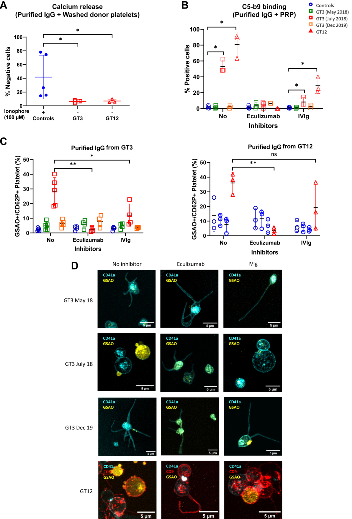
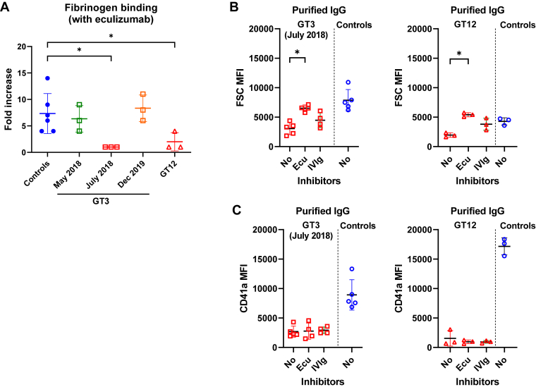
Results: Only 2 samples were able to severely block integrin function. We observed that these 2 sera caused a reduction in platelet size similar to that observed when platelets become procoagulant. Mixing healthy donor platelets with patients' sera or purified IgGs led to microvesiculation, phosphatidylserine exposure, and induction of calcium influx. This was associated with an increase in procoagulant platelets. Pore formation and calcium entry were associated with complement activation, leading to the constitution of a membrane attack complex (MAC) with enhanced complement protein C5b-9 formation. This process was inhibited by the complement 5 inhibitor eculizumab and reduced by polyvalent human immunoglobulins.

Conclusion: Our data suggest that complement activation induced by rare blocking anti-αIIbβ3 isoantibodies may lead to the formation of a MAC with subsequent pore formation, resulting in calcium influx and procoagulant platelet phenotype.

Keywords: Glanzmann thrombasthenia; anti-αIIbβ3 isoantibodies; coagulation; complement activation; platelet transfusion; procoagulant platelet.


Figures

Figure 1
Figure 1
(A) Inhibition of fibrinogen binding on platelet surface by patients’ sera. Different negative or positive sera from 12 patients with GT were incubated with control platelets for 20 minutes at 37 °C. Then, fibrinogen binding was analyzed by flow cytometry after activation with 10-μM ADP. Circles correspond to samples from GT3 (collected at different dates) and GT12. These samples were able to severely block Fg binding. Results obtained with different concentrations of abciximab (anti-αIIbβ3 MoAb) are also represented (positive control). Results were compared with those of the control group. Fibrinogen (B) and PAC-1 binding (C) measured by flow cytometry in GT3, GT12, and different day controls (n = 7-12). Activation was obtained by incubation with ADP (10 μM) for fibrinogen binding and TRAP-14 (50 μM) for PAC-1. Results were expressed as percentage of the MFI of controls. Mann–Whitney–Wilcoxon test (P value): ∗< .05, ∗∗< .01, and ∗∗∗∗< .0001. ADP, adenosine diphosphate; GT, Glanzmann thrombasthenia; MFI, mean fluorescence intensity.
Figure 2
Figure 2
(A) Dot plots of FSC (x axis) vs SSC fluorescence (y axis) show that GT3’s high positive serum (date: July 3, 2018) causes, at resting state, a change of platelet form similar to that observed when platelets are activated with calcium ionophore. (B) Histograms showing FSC values (MFI) obtained after mixing control platelets with GT3 (collected at different dates) and GT12’s sera. Also shown are results obtained with controls’ sera incubated with or without calcium ionophore (positive controls). (C, D) Analysis of PS exposure and MV formation by flow cytometry after incubation of controls’ citrated cPRP with controls, GT3, or GT12’s serum. Mann–Whitney–Wilcoxon test and Wilcoxon signed-rank test for controls incubated with ionophore or not. ∗< .05, ∗∗< .01, and ∗∗∗∗< .0001. cPRP, citrated platelet-rich plasma; FSC, forward scatter; MFI, mean fluorescence intensity; MV, microvesicle; PS, phosphatidylserine; SSC, side scatter.
Figure 3
Figure 3
Analysis of PS exposure (A) and MV formation (B) after incubation of cPRPs from 3 different patients with GT type I with sera of GT3, GT12, or controls incubated with ionophore (n = 5-6); nonretained flow-through (C) and IgG eluate fractions (D) were tested for their ability to induce PS exposure. Mann–Whitney–Wilcoxon test (P value): ∗< .05, ∗∗< .01, and ∗∗∗< .001. cPRP, citrated platelet-rich plasma; GT, Glanzmann thrombasthenia; IgG, immunoglobulin G; PS, phosphatidylserine.
Figure 4
Figure 4
(A) Purified IgGs from the patients or control donors (n = 3-13) were tested for their ability to induce calcium release. Also shown are control platelets incubated with ionophore as positive control. (B) Procoagulant platelets (GSAO+/CD62P+) induced by purified IgG from controls, GT3, or GT12’s serum on healthy donor platelets (n = 3-5). ANOVA with Tukey’s multiple comparisons test. (C) Confocal microscope images showing the procoagulant morphology of donor platelets stained with CD41a (BV510) and GSAO (Alexa-647) and treated with purified IgGs from GT3 or GT12. Platelet marker CD9 (Alexa-488) was included in samples treated with GT12 purified IgGs. (D) Nonlinear relationship between anti-αIIbβ3 antibody interference with CD41a binding (clone HIP8) and induction of procoagulant response in donor platelets (n = 3-5) treated with GT3- or GT12-purified IgG. Mann–Whitney–Wilcoxon test and Wilcoxon signed-rank test for controls incubated with ionophore or not. ∗< .05, ∗∗< .01, ∗∗∗< .001, and ∗∗∗∗< .0001. GT, Glanzmann thrombasthenia; IgG, immunoglobulin G.
Figure 5
Figure 5
(A) PS exposure from controls’ platelets pretreated with Src inhibitor PP2 (40 μM) or Rac1 inhibitor NSC23766 (300 μM) and eluates. Data are presented as median and interquartile. Mann–Whitney–Wilcoxon test P values: ∗< .05, ∗∗< .01. (B) Procoagulant platelets (GSAO+/CD62P+) induced by IgGs in the presence of FcγRIIa-blocking monoclonal antibody IV.3 (10 μg/mL). ANOVA with Tukey’s multiple comparisons test: ∗∗∗∗< .0001. IgG, immunoglobulin G; PS, phosphatidylserine.
Figure 6
Figure 6
(A) Purified IgGs from patients or control donors incubated with ionophore (100 μM) were tested for their ability to induce calcium release from washed control platelets (WP) in absence of a complement source. (B) Anti-αIIbβ3 isoantibodies induce the formation of a MAC, which was inhibited by eculizumab (50 μg/mL) or human polyclonal immunoglobulins (IVIg; 25 mg/mL). Formation of a MAC was measured employing an anti-C5b-9 antibody. (C) Control cPRP (n = 4-5) was pretreated with or without eculizumab (50 μg/mL) or human polyclonal immunoglobulins (IVIg; 25 mg/mL) before incubation with purified IgGs from GT3, GT12, or control donors and assayed by flow cytometry for procoagulant platelet response. Mixed effects analysis with Tukey’s multiple comparisons test: ∗< .05 and ∗∗< .01. (D) Confocal microscope imaging of donor platelets stained with CD41a (BV510), CD9 (Alexa-488), and GSAO (Alexa-647) and treated with purified IgGs from patients in the presence of eculizumab (50 micrograms/mL) or IVIg (25 mg/mL). IgG, immunoglobulin G; IVIg, intravenous immunoglobulin; MAC, membrane attack complex.
Figure 7
Figure 7
(A) Inhibition of fibrinogen binding on the platelet surface by anti-αIIbβ3 isoantibodies in the presence of eculizumab (50 μg/mL). (B) FSC and (C) CD41a median fluorescence intensity values obtained after mixing control platelets with GT3 collected at different dates, or GT12’s purified IgGs in the presence of eculizumab (50 μg/mL) in the presence of eculizumab (50 ug/mL) or human polyclonal immunoglobulins (IVIg, 25 mg/mL). Mann–Whitney–Wilcoxon test and Wilcoxon signed-rank test for controls incubated with ionophore or not. ∗< .05. FSC, forward scatter; IgG, immunoglobulin G; IVIg, intravenous immunoglobulin.

References

    1. Bledzka K., Smyth S.S., Plow E.F. Integrin αIIbβ3: from discovery to efficacious therapeutic target. Circ Res. 2013;112:1189–1200. - PMC - PubMed
    1. Nurden A.T. Platelet membrane glycoproteins: a historical review. Semin Thromb Hemost. 2014;40:577–584. - PubMed
    1. Botero J.P., Lee K., Branchford B.R., Bray P.F., Freson K., Lambert M.P., et al. Glanzmann thrombasthenia: genetic basis and clinical correlates. Haematologica. 2020;105:888–894. - PMC - PubMed
    1. Nurden A.T., Fiore M., Nurden P., Pillois X. Glanzmann thrombasthenia: a review of ITGA2B and ITGB3 defects with emphasis on variants, phenotypic variability, and mouse models. Blood. 2011;118:5996–6005. - PubMed
    1. Nurden A.T. Glanzmann thrombasthenia. Orphanet J Rare Dis. 2006;1:10. - PMC - PubMed

LinkOut - more resources