

Neurocysticercosis: A neglected but preventable cause of seizure in adults
- PMID: 38268622
- PMCID: PMC10805999
- DOI: 10.1002/ccr3.8454
Neurocysticercosis: A neglected but preventable cause of seizure in adults
Abstract
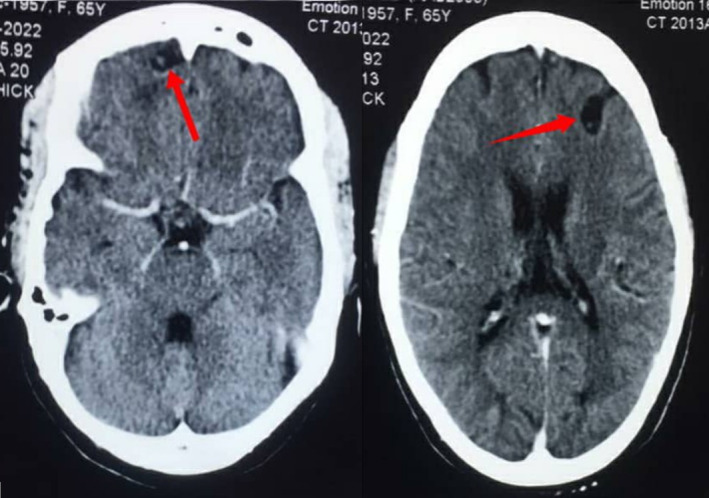
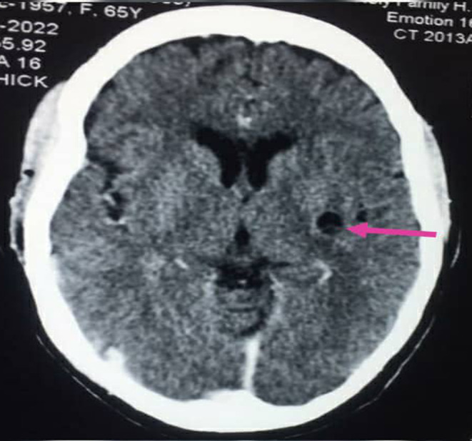
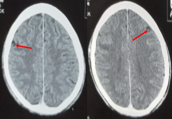
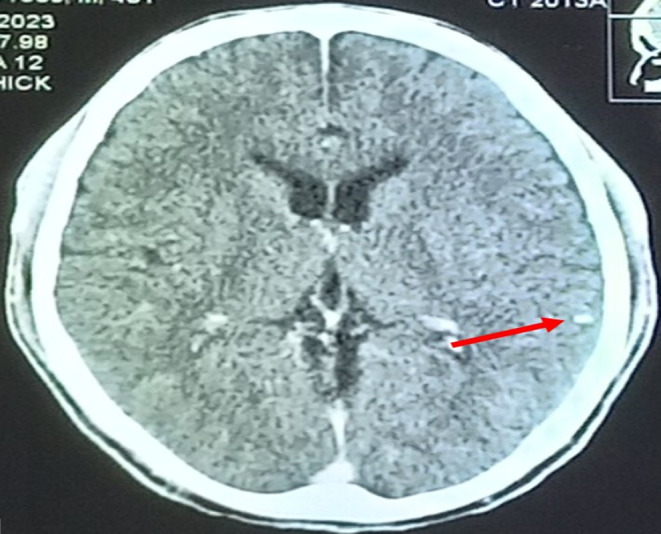
Neurocysticercosis is an infection of the central nervous system caused by the larval stage of Taenia solium. Although endemic in sub-Saharan Africa, it is neglected but remains a significant cause of preventable seizure in adults. Its diagnosis is challenging and is frequently missed due to its variable clinical manifestations and lack of diagnostic facilities in most areas of sub-Saharan Africa. This report discusses two cases of parenchymal neurocysticercosis in Ghanaians who presented to the emergency unit of a District Hospital with adult-onset seizures. The two cases highlight the need for a high index of suspicion and also underscore the important role of neuroimaging in the evaluation of patients presenting with adult-onset seizures in neurocysticercosis endemic areas. This is necessary for prompt detection and initiation of appropriate therapy in order to improve prognosis.
Keywords: Cysticercosis; Neurocysticercosis; Taenia solium; epilepsy; focal seizure; neuroimaging.
© 2024 The Authors. Clinical Case Reports published by John Wiley & Sons Ltd.
Conflict of interest statement
The authors declare no potential conflicts of interest with respect to the authorship and/or publication of this article.
Figures
References
-
- Singh G, Burneo JG, Sander JW. From seizures to epilepsy and its substrates: neurocysticercosis. Epilepsia. 2013;54:783‐792. - PubMed
-
- World Health Organization . WHO Guidelines on Management of Taenia solium Neurocysticercosis. 2021. - PubMed
-
- Kaur R, Arora N, Rawat SS, et al. Vaccine for a neglected tropical disease Taenia solium cysticercosis: fight for eradication against all odds. Expert Review of Vaccines. 2021;20:1447‐1458. - PubMed
-
- Owolabi LF, Adamu B, Jibo AM, Owolabi SD, Imam AI, Alhaji ID. Neurocysticercosis in people with epilepsy in sub‐Saharan Africa: a systematic review and meta‐analysis of the prevalence and strength of association. Seizure. 2020;76:1‐11. - PubMed
Publication types
LinkOut - more resources
Full Text Sources
