. 2023 Dec 26;27(1):108777.
doi: 10.1016/j.isci.2023.108777. eCollection 2024 Jan 19.

Rapamycin-sensitive mechanisms confine the growth of fission yeast below the temperatures detrimental to cell physiology

Affiliations

Rapamycin-sensitive mechanisms confine the growth of fission yeast below the temperatures detrimental to cell physiology

Yuichi Morozumi et al. iScience. .

Abstract

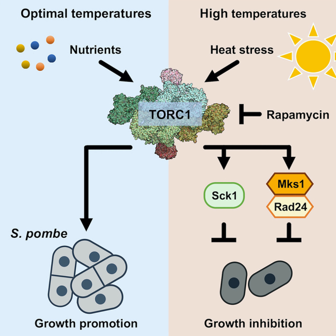
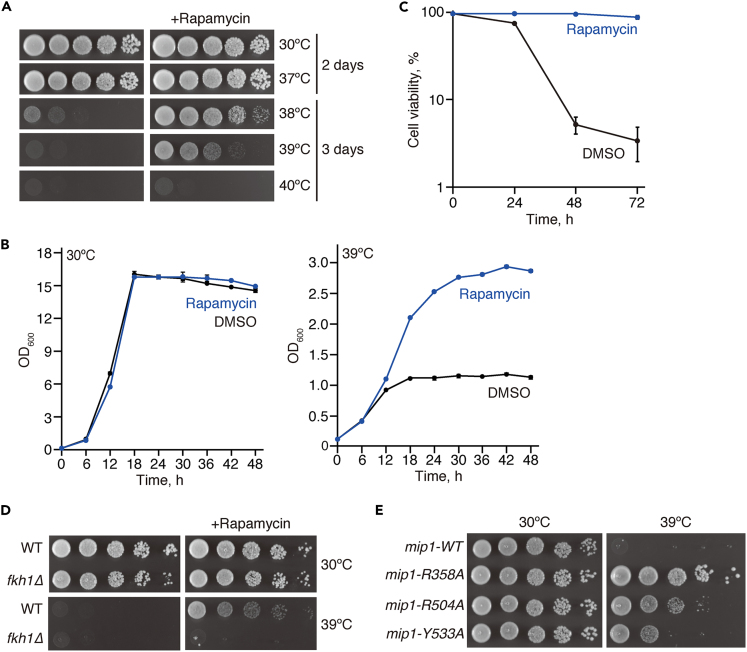
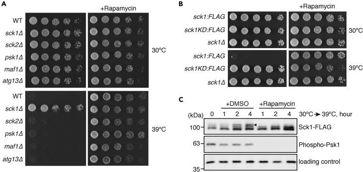
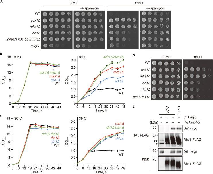
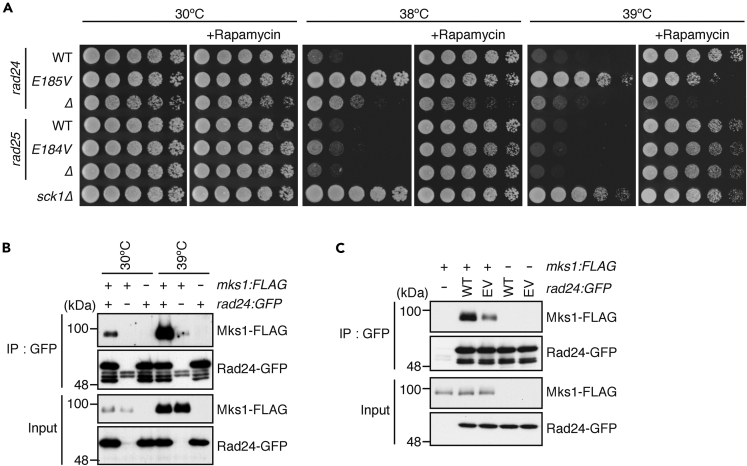
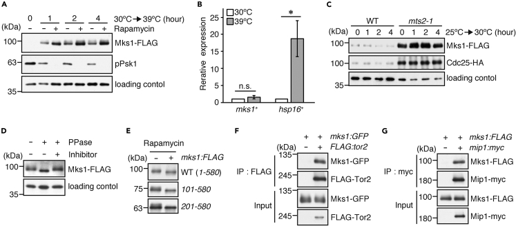
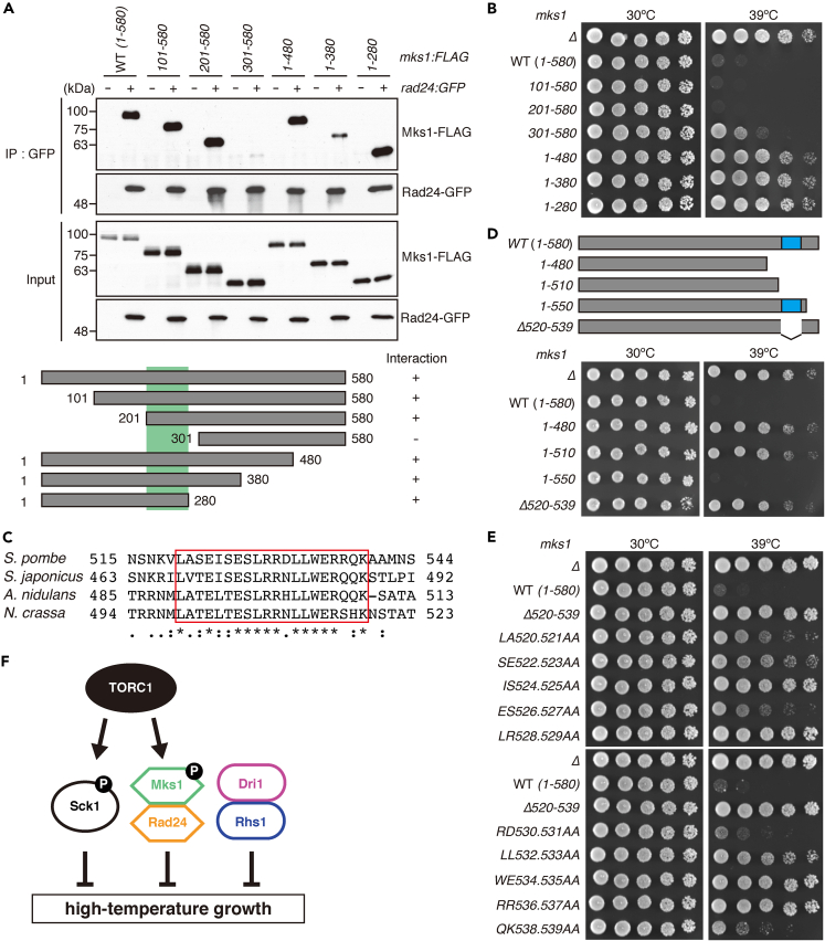
Cells cease to proliferate above their growth-permissible temperatures, a ubiquitous phenomenon generally attributed to heat damage to cellular macromolecules. We here report that, in the presence of rapamycin, a potent inhibitor of Target of Rapamycin Complex 1 (TORC1), the fission yeast Schizosaccharomyces pombe can proliferate at high temperatures that usually arrest its growth. Consistently, mutations to the TORC1 subunit RAPTOR/Mip1 and the TORC1 substrate Sck1 significantly improve cellular heat resistance, suggesting that TORC1 restricts fission yeast growth at high temperatures. Aiming for a more comprehensive understanding of the negative regulation of high-temperature growth, we conducted genome-wide screens, which identified additional factors that suppress cell proliferation at high temperatures. Among them is Mks1, which is phosphorylated in a TORC1-dependent manner, forms a complex with the 14-3-3 protein Rad24, and suppresses the high-temperature growth independently of Sck1. Our study has uncovered unexpected mechanisms of growth restraint even below the temperatures deleterious to cell physiology.

Keywords: Biological sciences; Microbial physiology; Microbiology; Molecular microbiology.


Conflict of interest statement

The authors declare no competing interests.

Figures

None
Graphical abstract
Figure 1
Figure 1
Suppression of TORC1 activity allows S. pombe cells to grow at high temperatures (A) Wild-type cells were grown in YES liquid medium at 30°C. Their serial dilutions were spotted onto YES agar plates in the presence and absence of 100 ng/mL rapamycin for growth assay at 30°C and 37°C for 2 days, and at 38°C, 39°C, and 40°C for 3 days. (B) Wild-type cells were grown in YES liquid medium at 30°C. At 0 h, the initial OD was adjusted to OD600 = 0.1 ± 0.02, and the cell growth in the presence and absence of 200 ng/mL rapamycin at 30°C and 39°C was monitored by measuring OD600 at indicated time points. Data are presented as means ± standard deviation (SD) from three independent experiments. (C) Wild-type cells were grown in YES liquid medium at 30°C. At time 0, the culture concentration was adjusted to OD600 = 0.1 ± 0.05, and the cell viability in the presence and absence of 200 ng/mL rapamycin at 39°C was measured by methylene blue staining at indicated time points. Data are presented as means ± SD from three independent experiments, and at least 200 cells were counted for each experiment. (D) The fkh1Δ mutant failed to grow at 39°C even in the presence of rapamycin. The growth of wild-type (WT) and fkh1Δ cells was tested as in (A). (E) The mip1 mutants exhibit cell proliferation at 39°C even in the absence of rapamycin. The indicated mip1 mutant strains were grown in YES medium at 30°C, and their serial dilutions were spotted onto YES agar plates for growth assay at 30°C and 39°C. See also Figure S2.
Figure 2
Figure 2
The Sck1 kinase inhibits cell growth at high temperatures (A) Wild-type (WT) and the indicated null mutant cells were grown in YES liquid medium at 30°C. Their growth in the presence and absence of rapamycin (100 ng/mL) was tested at 30°C and 39°C by spotting the serial dilutions of their cultures on YES agar plates. (B) The catalytic activity of Sck1 is required for suppressing high-temperature growth. The growth of the sck1:FLAG, sck1-K311A: FLAG, and sck1Δ strains was tested as in (A). (C) TORC1-dependent phosphorylation of Sck1 at high temperatures. The sck1:FLAG strain was grown in YES medium at 30°C and shifted to 39°C in the presence and absence of 200 ng/mL rapamycin. Cells were harvested at indicated time points, and their cell lysate was subjected to immunoblotting using anti-FLAG, anti-phospho-S6K1 (Phospho-Psk1) and anti-Spc1 (loading control) antibodies. An arrowhead represents the Sck1 protein phosphorylated by TORC1.
Figure 3
Figure 3
Identification of genes that suppress cellular growth at high temperatures (A) Isolated gene deletion mutants that exhibit growth at high temperatures. The indicated null mutants were grown in YES medium at 30°C. Their growth in the presence and absence of rapamycin (100 ng/mL) was tested at 30°C and 39°C by spotting serial dilutions of their cultures on YES agar plates. (B) Sck1 and Mks1 function independently to inhibit cell growth at high temperatures. Wild-type (WT) and the indicated mutant cells were cultured in YES medium at 30°C. At time 0, the culture concentration was adjusted to OD600 = 0.1 ± 0.02, and their growth at 30°C and 39°C was monitored by measuring OD600 every 6 h. Data are presented as means ± SD from three independent experiments. (C and D) Dri1 and Rhs1 function in the same pathway. (C) The growth kinetics of indicated mutant strains were measured as in (B). (D) The indicated mutant strains were grown in YES medium at 30°C, and their serial dilutions were spotted onto YES agar plates for growth assay at 30°C and 39°C. (E) Dri1 physically interacts with Rhs1. Strains expressing Rhs1-FLAG and Dri1-myc individually or simultaneously were grown in YES medium at 30°C and 39°C, and the cell lysate was subjected to immunoprecipitation using anti-FLAG affinity beads (IP: FLAG). The samples were analyzed by anti-FLAG and anti-myc immunoblotting. Single and double asterisks indicate immunoglobulin heavy chains and a non-specific protein recognized by the anti-FLAG antibody, respectively. See also Figures S3 and S4.
Figure 4
Figure 4
The rad24-E185V mutation, which compromises the Rad24-Mks1 interaction, confers heat resistance on S. pombe cells (A) The rad24-E185V mutation confers heat resistance on fission yeast cells. The indicated strains were cultured at 30°C. Their growth in the presence and absence of rapamycin (100 ng/mL) was tested at indicated temperatures by spotting serial culture dilutions on YES agar plates. (B) Mks1 physically interacts with Rad24. Strains expressing Mks1-FLAG and Rad24-GFP individually or simultaneously were grown in YES medium at 30°C and sifted to 39°C. After 2-h incubation at 39°C, the cell lysate was prepared, and Rad24-GFP was immunopurified using the antibodies against GFP (IP: GFP). Co-purified Mks1-FLAG was analyzed by immunoblotting. (C) The rad24-E185V mutation compromises the interaction of Rad24 with Mks1. mks1-FLAG cells expressing either Rad24 or Rad24-E185V tagged with GFP were grown, and their cell lysate was subjected to immunoprecipitation followed by immunoblotting as in (B). See also Figure S4 and Table S1.
Figure 5
Figure 5
Cellular Mks1 protein, which is phosphorylated by TORC1, accumulates upon heat stress by attenuation of its proteasomal degradation (A) Heat stress induces the accumulation of Mks1, which is further enhanced by rapamycin. Cells were grown in YES medium at 30°C and shifted to 39°C in the presence and absence of 200 ng/mL rapamycin. Their cell lysate was probed with anti-FLAG and anti-Spc1 (loading control) antibodies. (B) Heat stress does not significantly change the transcription of the mks1+ gene. The mRNA expression of mks1+ and hsp16+ was examined by qRT-PCR using cells grown in YES medium at 30°C and 39°C for 4 h. The expression level of each gene is presented as a value relative to that at 30°C. Bars indicate ± SD from two independent experiments in triplicate. ∗p < 0.05; n.s., not significant, compared to the mRNA expression at 30°C using Student’s t test. (C) The protein accumulation of Mks1 in the mts2-1 mutant. Wild-type (WT) and mts2-1 mutant cells were grown in YES medium at 25°C and shifted to 30°C. Cells were harvested at indicated time points, and their cell lysate was subjected to immunoblotting using anti-FLAG, anti-HA, and anti-Spc1 (loading control) antibodies. Cdc25, known to accumulate in the mts2-1 mutant, was utilized as a positive control. (D) Mks1 is a phosphoprotein. Cell lysate prepared from a strain expressing FLAG-tagged Mks1 was treated with lambda phosphatase (PPase) in the presence or absence of phosphatase inhibitor and subjected to immunoblotting as in (A). (E) Mks1 is phosphorylated in a manner dependent on TORC1. Cells expressing the indicated FLAG-tagged Mks1 proteins were grown in YES medium at 30°C and shifted to 39°C in the presence and absence of 200 ng/mL rapamycin. Their cell lysate was probed as in (A). Note that as rapamycin induces the Mks1 protein accumulation, the protein loading amount between rapamycin-treated and -untreated samples was adjusted to obtain comparable signals of Mks1. (F and G) Physical interaction between TORC1 and Mks1. Strains expressing Mks1-GFP and FLAG-Tor2 (F), or Mks1-FLAG and Mip1-myc (G) individually or simultaneously were grown in YES medium at 30°C and sifted to 39°C. After 2-h incubation at 39°C, FLAG-Tor2 (F) or Mip1-myc (G) was immunopurified from cell lysate, and co-purified Mks1 was analyzed by immunoblotting. See also Figure S5 and Tables S2–S4.
Figure 6
Figure 6
The C-terminal conserved region of Mks1 is indispensable for the suppression of high-temperature growth (A) The interaction of Rad24 with various truncated Mks1 fragments. rad24:GFP cells expressing various Mks1 fragments were cultured in YES medium at 30°C and sifted to 39°C. After 2-h incubation at 39°C, their cell lysate was subjected to immunoprecipitation using the anti-GFP antibodies (IP: GFP), and co-purified Mks1-FLAG was analyzed by immunoblotting. A green box in the schematic diagram of the Mks1 fragments indicates the region required for the Mks1-Rad24 interaction. (B) The growth of the strains expressing the various Mks1 fragments at high temperatures. The indicated mks1 mutant strains were grown in YES medium at 30°C, and their serial dilutions were spotted onto YES agar plates for growth assay at 30°C and 39°C. (C) Amino acid sequence alignment of the C-terminal conserved region of Mks1 in S. pombe, S. japonicus, A. nidulans, and N. crassa. The sequence alignment was performed using the CLUSTALW program (https://www.genome.jp/tools-bin/clustalw). Asterisks, identical amino acids; single and double dots, weakly and strongly similar amino acids, respectively. The conserved sequence stretches are shown in a red box. (D and E) The C-terminal conserved region of Mks1 is indispensable for its function of suppressing cell growth at high temperatures. The growth of the indicated mks1 mutant cells at 39°C was examined as in (B). A blue box in a schematic diagram of the Mks1 fragments in (D) indicates the conserved C-terminal region. (F) A model illustrating the negative regulation of cell growth under heat stress in fission yeast. See also Figure S7.

Similar articles

Cited by

References

    1. Mogk A., Bukau B., Kampinga H.H. Cellular Handling of Protein Aggregates by Disaggregation Machines. Mol. Cell. 2018;69:214–226. - PubMed
    1. Tyedmers J., Mogk A., Bukau B. Cellular strategies for controlling protein aggregation. Nat. Rev. Mol. Cell Biol. 2010;11:777–788. - PubMed
    1. Richter K., Haslbeck M., Buchner J. The Heat Shock Response: Life on the Verge of Death. Mol. Cell. 2010;40:253–266. - PubMed
    1. Verghese J., Abrams J., Wang Y., Morano K.A. Biology of the Heat Shock Response and Protein Chaperones: Budding Yeast (Saccharomyces cerevisiae) as a Model System. Microbiol. Mol. Biol. Rev. 2012;76:115–158. - PMC - PubMed
    1. Gidalevitz T., Prahlad V., Morimoto R.I. The stress of protein misfolding: From single cells to multicellular organisms. Cold Spring Harb. Perspect. Biol. 2011;3:a009704–a009718. - PMC - PubMed

LinkOut - more resources