. 2024 Mar;20(3):2262-2272.
doi: 10.1002/alz.13692. Epub 2024 Jan 25.

Down Syndrome Biobank Consortium: A perspective

Affiliations

Down Syndrome Biobank Consortium: A perspective

Iban Aldecoa et al. Alzheimers Dement. 2024 Mar.

Abstract

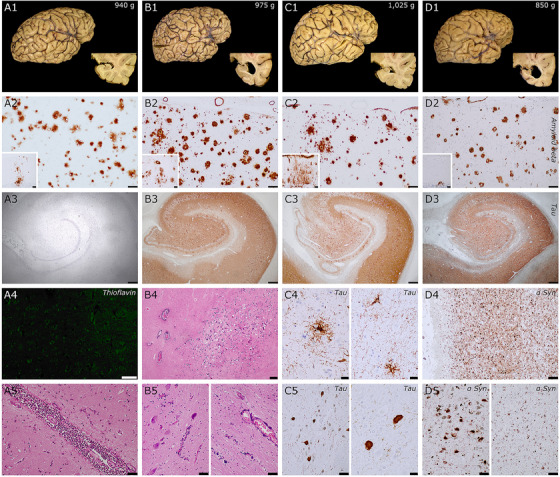
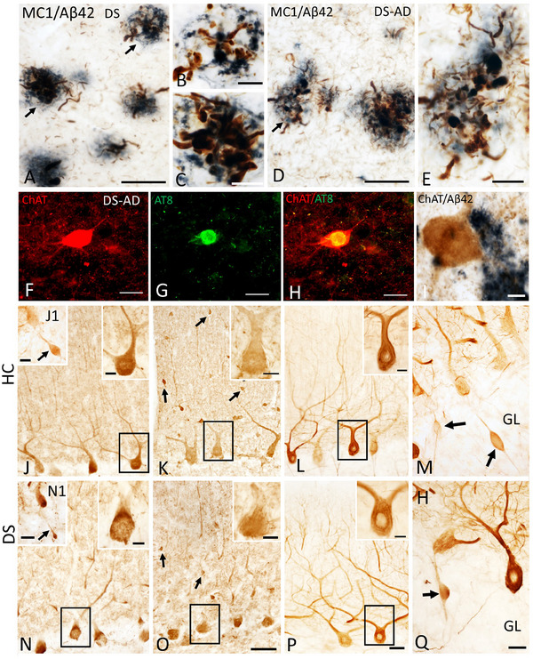
Individuals with Down syndrome (DS) have a partial or complete trisomy of chromosome 21, resulting in an increased risk for early-onset Alzheimer's disease (AD)-type dementia by early midlife. Despite ongoing clinical trials to treat late-onset AD, individuals with DS are often excluded. Furthermore, timely diagnosis or management is often not available. Of the genetic causes of AD, people with DS represent the largest cohort. Currently, there is a knowledge gap regarding the underlying neurobiological mechanisms of DS-related AD (DS-AD), partly due to limited access to well-characterized brain tissue and biomaterials for research. To address this challenge, we created an international consortium of brain banks focused on collecting and disseminating brain tissue from persons with DS throughout their lifespan, named the Down Syndrome Biobank Consortium (DSBC) consisting of 11 biobanking sites located in Europe, India, and the USA. This perspective describes the DSBC harmonized protocols and tissue dissemination goals.

Keywords: Alzheimer's disease; Down syndrome; biobanking; brain banking; repository; research.


Conflict of interest statement

The authors declare no conflicts of interest. Author disclosures are available in the supporting information.

Figures

FIGURE 1
FIGURE 1
Neuropathological features of four donors with DS. A1‐5 is a female in her mid 20s, B1‐5 a male in his mid 40s, C1‐5 a male over 70 years old, and D1‐5 a female in her mid 60s. DS donors B and D had an additional clinical diagnosis of Alzheimer's disease (AD). Similarities: First row: macroscopic features: all cases had low brain weight, regardless of neuropathological processes; donors with concomitant AD (B and D) had more severe hippocampal atrophy. Second row: all donors had abundant Aβ in the frontal cortex that extended to cerebellum (insets) in all cases, compatible with a Thal phase 5/5. Third row: A, younger donor, lacking tau pathology, while the three other donors had extensive neurofibrillary pathology in hippocampal complex (Braak stages IV, C3) ) and V/VI ( B3 and D3). Unique features. In donor A, despite extensive Aβ deposits, the case was almost devoid of neuritic plaques (A4, negative thioflavin staining in neocortex). This case also had acute meningitis, that was related to the final pathological processes associated with death (bottom row, A5, pericentral cortex). Case B had extensive calcium deposits in basal ganglia (B4, globus pallidum), dentate nucleus of the cerebellum (B5, bottom row), and adjacent white matter (B5), arteriolar walls and in pericapillary areas. This pattern is reminiscent of Fahr's syndrome, that was previously reported in cases with DS. Case C had an additional tauopathy compatible with progressive supranuclear palsy, with tufted astrocytes in motor cortex (C4 left) and putamen (C4 right) and neurofibrillary tangles in substantia nigra (C5 left), subthalamic nucleus (C5 right panel), globus pallidus. and additional features such as coiled bodies (not shown). Case D also had an extensive Lewy pathology, with severe involvement of the limbic system (D4, amygdala), brainstem (D5 left, substantia nigra) neocortex (D5 right, frontal cortex). Scale bars: A3 to D3 = 500 μm; B4 = 200 μm; A2 to D2, C5 left, D4, D5 = 100 μm; A2 to D2 insets, A4, A5, B5 = 50 μm; C4, C5 right = 20 μm.
FIGURE 2
FIGURE 2
Photomicrographs of dual labeled frontal cortex sections showing dystrophic neurites displaying immunoreactivity for tau conformational epitope MC1 (brown) intermingled within Aβ42‐ir plaques (blue) in a 47‐year‐old female nondemented (A) and a 46‐year‐old male demented (D) individual with DS. Note the presence of numerous MC1 immunoreactive (‐ir) neuropile threads in demented compared to nondemented DS case. (B, C, and E) High‐power images showing bulbous nature of dystrophic neurites within Aβ42‐ir plaques from panels A and D (arrows), respectively. (F–H) Single immunofluorescence images showing normal appearing striatal choline acetyltransferase (ChAT) positive neuron (red, F), AT8‐positive NFT (green; G) in a 46‐year‐old male demented case. (H) Merged image of ChAT and AT8 immunostaining shown in F and G.  Note the intact appearance of the cholinergic striatal neuron (red) despite the presence of an AT8 reactivity (yellow) within the perikarya in this demented DS case. (I) Intact ChAT‐positive putaminal neuron (brown) despite its proximity to Aβ42 staining (blue‐black) in a 46‐year‐old male donor with DS and dementia. (J, N) Photomicrographs showing Calb‐ir Purkinje cells (PCs) in a female 66‐year‐old healthy control (HC) (J) and a female 47‐year‐old nondemented DS (N) case. Upper right insets show high‐power image of black‐boxed Calb‐ir PCs in panels J and N.  Insets J1 and N1 show cerebellar granular layer (GL) Calb‐ir axonal torpedoes (arrows) in a male 51‐year‐old healthy control (HC) and a female nondemented 60‐year‐old DS case. (K, O) Images showing Parv‐ir PCs and Parv‐ir interneurons (black arrows) within the cerebellar molecular layer (ML) in a female 69‐year‐old HC (K) and a female 44‐year‐old DS without dementia (O). Upper right insets (K, O) are higher‐magnification images of the Parv‐ir PCs shown in the black boxes. (L, P). Photomicrographs of nonphosphorylated high‐molecular‐weight neurofilaments (SMI‐32‐ir) PC dendritic arbors and axons in a female 69‐year‐old HC (L) and a male 46‐year‐old DS‐AD (P) case. Insets in L and P show high‐power images of boxed SMI‐32‐ir PCs and proximal dendrites. (M, Q) Swollen SMI‐32‐ir proximal PC axons or torpedoes (arrows) in GL of male 51‐year‐old HC (M) and female 60‐year‐old DS (Q). Scale bars:  A, D, F–H = 50 μm; B, C, E, I and insets in J, K, L, N, O, P = 10 μm; J1 and N1 insets = 30 μm; O = 50 μm and applies to J, K, M; P = 50 μm and applies to L; Q = 25 μm and applies to M.

Similar articles

Cited by

References

    1. Zigman WB. Atypical aging in Down syndrome. Dev Disabil Res Rev. 2013;18:51‐67. - PubMed
    1. Hartley D, Blumenthal T, Carrillo M, et al. Down syndrome and Alzheimer's disease: common pathways, common goals. Alzheimers Dement. 2015;11:700‐709. - PMC - PubMed
    1. Wisniewski KE, Dalton AJ, McLachlan C, Wen GY, Wisniewski HM. Alzheimer's disease in Down's syndrome: clinicopathologic studies. Neurology. 1985;35:957‐961. - PubMed
    1. Wisniewski KE, Wisniewski HM, Wen GY. Occurrence of neuropathological changes and dementia of Alzheimer's disease in Down's syndrome. Ann Neurol. 1985;17:278‐282. - PubMed
    1. Iulita MF, Garzon Chavez D, Klitgaard Christensen M, et al. Association of Alzheimer disease with life expectancy in people with Down syndrome. JAMA Netw Open. 2022;5:e2212910. - PMC - PubMed

Publication types