. 2024 Aug;17(4):768-778.
doi: 10.1007/s12265-024-10482-1. Epub 2024 Jan 25.

Dysregulation of micro-RNA 143-3p as a Biomarker of Carotid Atherosclerosis and the Associated Immune Reactions During Disease Progression

Affiliations

Dysregulation of micro-RNA 143-3p as a Biomarker of Carotid Atherosclerosis and the Associated Immune Reactions During Disease Progression

Paula González-López et al. J Cardiovasc Transl Res. 2024 Aug.

Abstract

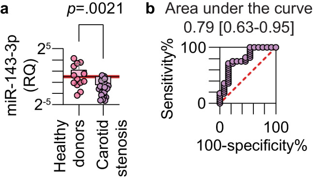
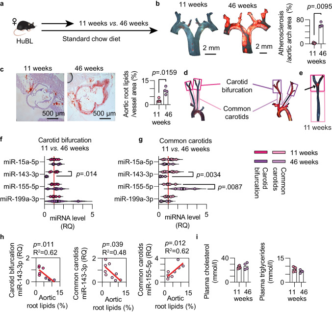
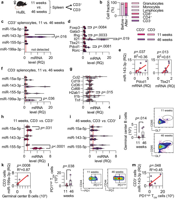
Atherosclerosis commonly remains undiagnosed until disease manifestations occur. The disease is associated with dysregulated micro(mi)RNAs, but how this is linked to atherosclerosis-related immune reactions is largely unknown. A mouse model of carotid atherosclerosis, human APOB100-transgenic Ldlr-/- (HuBL), was used to study the spatiotemporal dysregulation of a set of miRNAs. Middle-aged HuBL mice with established atherosclerosis had decreased levels of miR-143-3p in their carotid arteries. In young HuBL mice, early atherosclerosis was observed in the carotid bifurcation, which had lower levels of miR-15a-5p, miR-143-3p, and miR-199a-3p, and higher levels of miR-155-5p. The dysregulation of these miRNAs was reflected by specific immune responses during atheroprogression. Finally, levels of miR-143-3p were 70.6% lower in extracellular vesicles isolated from the plasma of patients with carotid stenosis compared to healthy controls. Since miR-143-3p levels progressively decrease when transitioning between early and late experimental carotid atherosclerosis, we propose it as a biomarker for atherosclerosis.

Keywords: Atherosclerosis; Carotid stenosis; Dyslipidemia; Micro-RNA; T-lymphocytes.


Figures

Fig. 1
Fig. 1
Plasma detection of miR-143-3p in carotid stenosis patients. (a) miRNAs in extracellular vesicles were isolated from the plasma of patients with advanced atherosclerosis and the plasma from healthy donors without manifest atherosclerosis. The relative expression of miR-143-3p was measured by real-time PCR (Mann-Whitney test, healthy donors n = 13, carotid stenosis n = 28, log2-scaled y-axis) (b) A receiver operating characteristic curve analysis illustrates how miR-143-3p can be used as a biomarker for carotid atherosclerosis
Fig. 2
Fig. 2
Dysregulation of a set of miRNAs in mouse carotid arteries. (a) Experimental setup comparing atheroprogression in HuBL mice. (b) Micrographs of Sudan-IV-stained aortic arches with a 2 mm scale bar and quantification of the atherosclerotic plaques (orange color) divided by total aortic arch area. (c) Micrographs of Oil Red O-stained sections 300 µm from the aortic root with a 500 µm scale bar. Mean aortic root lipids were quantified (red color) and divided by aortic cross-section area. (d) Sudan-IV stained aortic arches with branches to depict microdissection of common carotids and carotid bifurcation. (e) Micrograph of a carotid artery from an 11-week-old HuBL mouse, the arrow indicates Sudan-IV+ plaque in the carotid bifurcation. (f-g) Relative quantification of miRNAs in the carotid bifurcation (bright colors) and common carotids (pale colors) from the 11-week (pink colors, n = 6) and 46-week (purple colors, n = 4) old female HuBL mice. (h) Linear regression between lipid deposition in the aortic root and the levels of miR-143-3p in the carotid bifurcation and between lipid deposition in the aortic root and the levels of miR-155-5p and miR-143-3p in the common carotids. (i) Plasma concentration of cholesterol and triglycerides
Fig. 3
Fig. 3
Site-specific miRNA level changes in early and late carotid atherosclerosis. (a-b) Relative quantification of miRNAs in the common carotids (pale colors) and carotid bifurcation (bright colors) from the 11-week (pink colors, n = 6) and 46-week (purple colors, n = 4) old female HuBL mice. (c) mRNA levels for genes of interest in 11-week-old mice. (d) Linear regression between Ccl2 mRNA and miR-155-5p (left) and miR-199a-3p (right) in carotids with early atherosclerosis. (e) mRNA levels for genes of interest in 46-week-old mice. (f) Linear regression between Cd68 mRNA (left) and Vcam1 mRNA (right), respectively, and miR-143-3p in carotids with advanced atherosclerosis
Fig. 4
Fig. 4
miRNA levels in splenic immune cells during atheroprogression. (a) Experimental setup of CD3+ and CD3 cell isolation. (b) Cell fractions before and after isolation of CD3+ cells from the spleen. (c) miRNA levels in CD3+ cells from the 11-week (pink, n = 6) and 46-week (purple, n = 4) old female HuBL mice. (d) mRNA levels for genes of interest in CD3+ cells. (e) Linear regression between miR-143-3p in CD3+ cells and Pdcd1 and Tbx21 mRNA. (f) miRNA levels in CD3 cells. (g) mRNA levels in CD3 cells. (h-i) miRNA levels in the CD3+ and CD3 cells from the 11- and 46-week groups. (j-k) Germinal center B cells as quantified using flow cytometry from the mediastinal and renal lymph nodes and their correlation to miR-199a-3p in splenic CD3 cells. (l-m) PD1high TEM cells in the mediastinal and renal lymph nodes and their correlation to miR-143-3p in splenic CD3+ cells
Fig. 5
Fig. 5
Dysregulated transcripts in iliac lymph nodes of middle-aged mice. (a) Experimental setup. (b) Micrographs of Sudan-IV-stained aortas with a 4 mm scale bar. (c) Total plasma cholesterol and triglycerides concentrations. (d) Relative quantification of miRNAs in the iliac lymph nodes from male Ldlr−/ (pink color, n = 8) and HuBL mice (purple color, n = 8). (e–f) Linear regression between miR-15a-5p and miR-199-3p with plasma cholesterol and triglycerides. (g) mRNA levels for genes of interest in the iliac lymph nodes. (h) Linear regression between Pdcd1 and Ifng mRNA, respectively, and miR-15a-5p levels in the iliac lymph nodes

References

    1. Gisterå A, Hansson GK. The immunology of atherosclerosis. Nat Rev Nephrol. 2017;13(6):368–80. 10.1038/nrneph.2017.51. 10.1038/nrneph.2017.51 - DOI - PubMed
    1. Chèvre R, González-Granado JM, Megens RT, Sreeramkumar V, Silvestre-Roig C, Molina-Sánchez P, et al. High-resolution imaging of intravascular atherogenic inflammation in live mice. Circ Res. 2014;114(5):770–9. 10.1161/circresaha.114.302590. 10.1161/circresaha.114.302590 - DOI - PubMed
    1. Libby P. The changing landscape of atherosclerosis. Nature. 2021;592(7855):524–33. 10.1038/s41586-021-03392-8. 10.1038/s41586-021-03392-8 - DOI - PubMed
    1. Ji R, Cheng Y, Yue J, Yang J, Liu X, Chen H, et al. MicroRNA expression signature and antisense-mediated depletion reveal an essential role of MicroRNA in vascular neointimal lesion formation. Circ Res. 2007;100(11):1579–88. 10.1161/circresaha.106.141986. 10.1161/circresaha.106.141986 - DOI - PubMed
    1. Elia L, Quintavalle M, Zhang J, Contu R, Cossu L, Latronico MV, et al. The knockout of miR-143 and -145 alters smooth muscle cell maintenance and vascular homeostasis in mice: correlates with human disease. Cell Death Differ. 2009;16(12):1590–8. 10.1038/cdd.2009.153. 10.1038/cdd.2009.153 - DOI - PMC - PubMed

Publication types

MeSH terms

LinkOut - more resources