. 2024 Feb 19;7(2):853-862.
doi: 10.1021/acsabm.3c00872. Epub 2024 Jan 25.

Reusable Colorimetric Biosensors on Sustainable Silk-Based Platforms

Affiliations

Reusable Colorimetric Biosensors on Sustainable Silk-Based Platforms

Augusto Márquez et al. ACS Appl Bio Mater. .

Abstract

In biosensor development, silk fibroin is advantageous for providing transparent, flexible, chemically/mechanically stable, biocompatible, and sustainable substrates, where the biorecognition element remains functional for long time periods. These properties are employed here in the production of point-of-care biosensors for resource-limited regions, which are able to display glucose levels without the need for external instrumentation. These biosensors are produced by photopatterning silk films doped with the enzymes glucose oxidase and peroxidase and photoelectrochromic molecules from the dithienylethene family acting as colorimetric mediators of the enzymatic reaction. The photopatterning results from the photoisomerization of dithienylethene molecules in the silk film from its initial uncolored opened form to its pink closed one. The photoisomerization is dose-dependent, and colored patterns with increasing color intensities are obtained by increasing either the irradiation time or the light intensity. In the presence of glucose, the enzymatic cascade reaction is activated, and peroxidase selectively returns closed dithienylethene molecules to their initial uncolored state. Color disappearance in the silk film is proportional to glucose concentration and used to distinguish between hypoglycemic (below 4 mM), normoglycemic (4-6 mM), and hyperglycemic levels (above 6 mM) by visual inspection. After the measurement, the biosensor can be regenerated by irradiation with UV light, enabling up to five measurement cycles. The coupling of peroxidase activity to other oxidoreductases opens the possibility to produce long-life reusable smart biosensors for other analytes such as lactate, cholesterol, or ethanol.

Keywords: colorimetric biosensor; dithienylethene mediators, photopatterning; glucose detection; photoelectrochromic; point of care; silk fibroin.


Conflict of interest statement

The authors declare no competing financial interest.

Figures
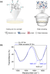
Figure 1
Figure 1
Formation of crystalline enzymatic silk films doped with DTE. (a) Illustration of the crystallization protocol, involving casting, drying, and water annealing in the vacuum. (b) ATR-FTIR spectra of dried (black) and crystalline silk films (blue).
Figure 2
Figure 2
Enzymatic silk films with photochromic activity. (a) Isomerization reaction of DTE molecules. The ring opening/closing of the structure is accompanied by a color change from transparent to pink when irradiated with UV. The reaction is reversible using visible light. (b) Immobilized DTE in enzymatic silk films containing GOx and HRP keeps the photochromism. The pads were placed over the printed word “Transparent” to denote the transparency of the film.
Figure 3
Figure 3
DTE cycle in enzymatic silk films. (a) Enzymatic cascade reaction where DTE acts as a reversible colorimetric mediator activated via UV light in the enzymatic silk films. SH represents a protic solvent, and S represents the corresponding radical species. (b) Nonirradiated doped SF pads (i) were exposed to UV for 30 s to close the DTE molecule resulting in the change of color (ii). One of the pads was maintained dry as a control, while a drop of PBS solution and a drop of 4 mM glucose PBS solution were added on top of the others (iii). After 10 min, only the pad in contact with the glucose solution lost the color. (c) Color of the enzymatic silk films was restored after a second UV irradiation in all samples. This process could be repeated a minimum of five times.
Figure 4
Figure 4
Photolithographic patterning of enzymatic silk films doped with DTE. (a) Photopatterning steps on SF functionalized with DTE, GOx, and HRP. After the first UV pattering, the same pattern is developed again on the film after turning the mask 90°. (b) Photographs of 4 mm-diameter SF films doped with DTE and enzymes after 30 s of UV irradiation using a photomask with straight lines. (c) It is possible to develop figures or regular patterns on the films using a suitable mask during the UV irradiation. (d) Photograph of 4 mm-diameter SF films after two consecutive irradiation steps with the same photomask, after shifting the photomask 90° in the second irradiation step.
Figure 5
Figure 5
Glucose detection using patterned enzymatic silk films. (a) First, a doped SF pad of 4 mm diameter is irradiated with UV for 30 s, using an aluminum mask to generate patterns of 200 μm. Then, the pad is incubated in PBS aqueous solution for 1 min with 0, 4, and 8 mM glucose. Finally, the pad is removed from the solution, dried, and irradiated using the same mask forming 90° angle with respect to the previous pattern. (b) On the left, microscopy images of silk pads in contact with different glucose solutions in PBS (0, 4, and 8 mM from top to bottom). The glucose PBS solution led to different color decays within the film. By image spectral analysis (on the right), it is possible to quantify the decay in different areas of the film: never irradiated area (black), irradiated area and incubated in 0, 4, or 8 mM glucose solution (red), irradiated area after solution removal (blue), and irradiated area before and after the immersion (purple).
Figure 6
Figure 6
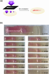
Gradient biosensor production and performance. (a) Patterning was done using a mask with a gradient of opacity to UV light. For 30 s, the SF enzymatic film was irradiated and a gradient of color was revealed on the surface. (b) Images corresponding to a sample photopatterned following procedure (a) and placed in contact with a 4 mM glucose solution in PBS for 14 min. It can be noted that the bleaching color takes places gradually from the less-intense colored side to the most intense along time.
Figure 7
Figure 7
Quantification of glucose levels using a photopatterned silk film display. (a) Scheme with the photolithographic procedure for the production of colorimetric silk displays enabling glucose levels quantification. Each rectangle is irradiated with increasing doses of UV light (from left to right). (b) Color of each rectangle after the irradiation doses was analyzed using ImageJ software. After splitting the image by colors, the green channel was selected for being the one providing the best sensibilities. (c) Eventually, four films were patterned identically and used to sense different concentrations of glucose in PBS (0, 2, 4, and 6 mM). The picture shows the color variation after 30 min on the displays. In pink, the last rectangle observable with the naked eye is shown. (d) Graph shows the bleaching color of each rectangle after the incubation in glucose solutions. Images were analyzed with ImageJ following the previous procedure. The green dashed line indicates the color intensity (0.1 au) at which it is possible to detect the bleaching color on the display.

References

    1. Land K. J.; Boeras D. I.; Chen X.-S.; Ramsay A. R.; Peeling R. W. REASSURED Diagnostics to Inform Disease Control Strategies, Strengthen Health Systems and Improve Patient Outcomes. Nat. Microbiol. 2019, 4 (1), 46–54. 10.1038/s41564-018-0295-3. - DOI - PMC - PubMed
    1. Meggi B.; Bollinger T.; Mabunda N.; Vubil A.; Tobaiwa O.; Quevedo J. I.; Loquiha O.; Vojnov L.; Peter T. F.; Jani I. V. Point-Of-Care P24 Infant Testing for HIV May Increase Patient Identification despite Low Sensitivity. PLoS One 2017, 12 (1), e016949710.1371/journal.pone.0169497. - DOI - PMC - PubMed
    1. Rei Yan S. L.; Wakasuqui F.; Wrenger C. Point-of-Care Tests for Malaria: Speeding up the Diagnostics at the Bedside and Challenges in Malaria Cases Detection. Diagn. Microbiol. Infect. Dis. 2020, 98 (3), 11512210.1016/j.diagmicrobio.2020.115122. - DOI - PubMed
    1. Mabey D. C.; Sollis K. A.; Kelly H. A.; Benzaken A. S.; Bitarakwate E.; Changalucha J.; Chen X.-S.; Yin Y.-P.; Garcia P. J.; Strasser S.; Chintu N.; Pang T.; Terris-Prestholt F.; Sweeney S.; Peeling R. W. Point-of-Care Tests to Strengthen Health Systems and Save Newborn Lives: The Case of Syphilis. PLoS Med. 2012, 9 (6), e100123310.1371/journal.pmed.1001233. - DOI - PMC - PubMed
    1. Chen X.-S. Rapid Diagnostic Tests for Neurosyphilis. Lancet Infect. Dis. 2013, 13 (11), 918–919. 10.1016/S1473-3099(13)70293-7. - DOI - PubMed

Publication types

LinkOut - more resources