. 2024 Mar 14;120(3):286-300.
doi: 10.1093/cvr/cvae025.

Ecto-CD38-NADase inhibition modulates cardiac metabolism and protects mice against doxorubicin-induced cardiotoxicity

Affiliations

Ecto-CD38-NADase inhibition modulates cardiac metabolism and protects mice against doxorubicin-induced cardiotoxicity

Thais R Peclat et al. Cardiovasc Res. .

Abstract

Aims: Doxorubicin (DXR) is a chemotherapeutic agent that causes dose-dependent cardiotoxicity. Recently, it has been proposed that the NADase CD38 may play a role in doxorubicin-induced cardiotoxicity (DIC). CD38 is the main NAD+-catabolizing enzyme in mammalian tissues. Interestingly, in the heart, CD38 is mostly expressed as an ecto-enzyme that can be targeted by specific inhibitory antibodies. The goal of the present study is to characterize the role of CD38 ecto-enzymatic activity in cardiac metabolism and the development of DIC.

Methods and results: Using both a transgenic animal model and a non-cytotoxic enzymatic anti-CD38 antibody, we investigated the role of CD38 and its ecto-NADase activity in DIC in pre-clinical models. First, we observed that DIC was prevented in the CD38 catalytically inactive (CD38-CI) transgenic mice. Both left ventricular systolic function and exercise capacity were decreased in wild-type but not in CD38-CI mice treated with DXR. Second, blocking CD38-NADase activity with the specific antibody 68 (Ab68) likewise protected mice against DIC and decreased DXR-related mortality by 50%. A reduction of DXR-induced mitochondrial dysfunction, energy deficiency, and inflammation gene expression were identified as the main mechanisms mediating the protective effects.

Conclusion: NAD+-preserving strategies by inactivation of CD38 via a genetic or a pharmacological-based approach improve cardiac energetics and reduce cardiac inflammation and dysfunction otherwise seen in an acute DXR cardiotoxicity model.

Keywords: CD38; Cardiotoxicity; DIC; Doxorubicin; Ecto-NADase; Heart failure; NAD+.


Conflict of interest statement

Conflict of interest: E.N.C. holds a patent on the use of CD38 inhibitors for metabolic diseases licensed by Elysium Health. E.N.C. is a consultant for TeneoBio, Calico, Mitobridge, and Cytokinetics. E.N.C. is on the advisory board of Eolo Pharma. E.N.C. and WVS own stocks in TeneoBio. Research in the E.N.C. laboratory has been conducted in compliance with Mayo Clinic’s conflict of interest policies.

Figures

None
This image was created in BioRender.com.
Figure 1
Figure 1
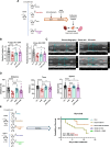
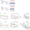
Genetic inactivation of CD38 catalytic activity protects mice against DIC. Adult (1-year-old) WT and CD38-CI mice were injected with a single dose of either 15 mg/kg of DXR or vehicle. (A) Experiment scheme showing all tests, imaging, and total duration of the experiment (n = 4–15 animals per group). (B) NAD+ levels and (C) CD38 activity in heart tissue from WT or CD38-CI mice treated with vehicle or DXR. (D) EF and FS were assessed by echocardiography. (E) Representative images from the short axis, M-mode view of echocardiography for each group. (F) Uphill treadmill exhaustion test performed 9 days after DXR injection. Graphs show running distance (m), time running (s), and maximal speed (m/s). (G) Body weight change 10 days after DRX treatment. All data are expressed as mean ± SEM, analysed by an unpaired two-sided t-test. *P < 0.05, **P < 0.01, ***P < 0.001, ****P < 0.0001.
Figure 2
Figure 2
CD38 plays a role in DIC and treatment with an anti-NADase-specific and non-cytotoxic antibody protects mice from DXR-induced cardiac dysfunction and overall mortality. Adult (1-year-old) WT mice that were injected with a single dose of either vehicle, 15 mg/kg of DXR, 5 mg/kg of Ab68, or 15 mg/kg of DXR and 5 mg/kg of Ab68 once before and twice after the DXR injection. (A) Experiment scheme showing all tests, imaging, and total duration of the experiment (n = 6–15 animals per group). (B) EF and FS were assessed by echocardiography 4 days after DXR injection. (C) Representative images from the short axis, M-mode view of echocardiography for each group. (D) Uphill treadmill exhaustion test performed after 9 days of DXR injection. Graphs show running distance (m), time running (s), and maximal speed (m/s). (E) Left panel—experiment scheme. Right panel—survival curve in days after the different treatments. All data are expressed as mean ± SEM, analysed by an unpaired two-sided t-test. The survival curve was analysed with a log-rank test and is represented in a Kaplan–Meier. *P < 0.05, **P < 0.01, ***P < 0.001, ****P < 0.0001.
Figure 3
Figure 3
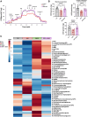
Treatment with Ab68 protects mice against DXR-induced mitochondrial dysfunction and improves the energetic metabolites supply in the heart. Adult (1-year-old) WT mice that were injected with a single dose of either vehicle, 15 mg/kg of DXR, 5 mg/kg of Ab68, or 15 mg/kg of DXR, and 5 mg/kg of Ab68 once before and twice after the DXR injection. (A and B) Respirometry analysis was performed in cardiac tissue from mice treated or not with DXR and Ab68. (A) Representative oxygraphy trace showing the oxygen consumption rate (OCR) and all substrates used during the experiment. (B) Maximal respiration, Complex I-dependent maximal respiration, and State 3 respiration. (C–E) Metabolomics analysis was detected using LC/MS/MS and Polar LC in cardiac tissues from WT mice treated or not with DXR and Ab68 (n = 6–8 animals per group). The data were analysed using MetaboAnalyst Software 5.0. (C) Heatmap showing the relative abundance of metabolites detected in each group (DXR, Ab68, and DXR + Ab68) compared with control and represented with colour intensity in which warmer colours (red/positive values) indicate higher values (in this case up-regulation) and colder colours (blue/negative values) indicate lower values (in this case down-regulation); (D) NAD+ precursors; (E) ADP ribose and citric acid metabolites. (F) Eicosanoids 12-HETE and 14-HDoHE/17-HDoHE. All data are expressed as mean ± SEM, analysed by an unpaired two-sided t-test. *P < 0.05, **P < 0.01, ***P < 0.001, ****P < 0.0001.
Figure 3
Figure 3
Treatment with Ab68 protects mice against DXR-induced mitochondrial dysfunction and improves the energetic metabolites supply in the heart. Adult (1-year-old) WT mice that were injected with a single dose of either vehicle, 15 mg/kg of DXR, 5 mg/kg of Ab68, or 15 mg/kg of DXR, and 5 mg/kg of Ab68 once before and twice after the DXR injection. (A and B) Respirometry analysis was performed in cardiac tissue from mice treated or not with DXR and Ab68. (A) Representative oxygraphy trace showing the oxygen consumption rate (OCR) and all substrates used during the experiment. (B) Maximal respiration, Complex I-dependent maximal respiration, and State 3 respiration. (C–E) Metabolomics analysis was detected using LC/MS/MS and Polar LC in cardiac tissues from WT mice treated or not with DXR and Ab68 (n = 6–8 animals per group). The data were analysed using MetaboAnalyst Software 5.0. (C) Heatmap showing the relative abundance of metabolites detected in each group (DXR, Ab68, and DXR + Ab68) compared with control and represented with colour intensity in which warmer colours (red/positive values) indicate higher values (in this case up-regulation) and colder colours (blue/negative values) indicate lower values (in this case down-regulation); (D) NAD+ precursors; (E) ADP ribose and citric acid metabolites. (F) Eicosanoids 12-HETE and 14-HDoHE/17-HDoHE. All data are expressed as mean ± SEM, analysed by an unpaired two-sided t-test. *P < 0.05, **P < 0.01, ***P < 0.001, ****P < 0.0001.
Figure 4
Figure 4
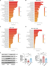
The anti-NADase Ab68 promotes metabolic shift, improving cardiac energetic metabolism. (A–D) Metabolomics analysis was detected using LC/MS/MS and Polar LC in cardiac tissues from WT mice treated or not with DXR and Ab68 (n = 6–8 animals per group). Graphs show the main biochemical pathways affected by DXR, Ab68, or both. (E) Representatives immunoblot and graphs showing levels of P-AMPK, AMPK, P-p70S6K, and P-70S6K with GADPH used as the loading control. All data are expressed as mean ± SEM, analysed by an unpaired two-sided t-test. *P < 0.05, **P < 0.01.
Figure 5
Figure 5
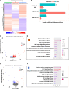
Ab68 reverses the inflammatory gene response induced by DXR treatment. (A) Heatmap of hierarchical clustering of DEGs among Ab68, DXR, and DXR + Ab68 treatment groups (X-axis represents each comparing sample. Y-axis represents DEGs. Colouring indicates the log2-transformed fold change; high, red; low, blue). (B) Bar plot showing the number of DEGs in each treatment group. (C) Volcano plot showing the up- and down-regulated genes by DXR treatment in comparison with the control group. (D) Lollipop plot showing the enriched up-regulated pathways by DXR treatment on the KEGG gene data set. (E) Volcano plot showing the up- and down-regulated genes in DXR + Ab68-treated mice in comparison with the DXR group. (F) Lollipop plot showing the enriched down-regulated pathways by Ab68 treatment in DXR-treated mice on the KEGG gene data set.
Figure 6
Figure 6
The anti-NADase Ab68 does not affect the anti-neoplastic effect of DXR. Six- to nine-week-old BALB/c mice were subcutaneously inoculated on the right flank with EMT-6 (breast cancer) or H22 (liver cancer) tumour cells. (A) Experiment schemes. For the EMT-6 experiment (upper), animals were divided into four groups: vehicle, DXR, Ab68, and DXR + Ab68. Animals received twice-a-week injections of either vehicle, 4 mg/kg of DXR, 5 mg/kg of Ab68, or both drugs. For the H22 experiment (lower), animals were divided into three groups: vehicle, DXR, and DXR + Ab68. Animals received twice-a-week injections of either vehicle, 5 mg/kg of DXR, or 5 mg/kg of DXR, and 5 mg/kg of Ab68 together. (B) EMT-6 experiment tumour size—L to R: %T/C, change in tumour volume over time, and final tumour volume on Day 21. (C) H22 experiment tumour size—L to R: %T/C, change in tumour volume over time, and final tumour volume on Day 13. All data are expressed as mean ± SEM, analysed by an unpaired two-sided t-test. Timeframe graphs showing tumour changes overtime were analysed with two-way ANOVA. *P < 0.05, **P < 0.01, ***P < 0.001, ****P < 0.0001. T/C: The T/C value (%) is an indicator of tumour response to treatment, where T and C are the mean tumour volumes of the treatment and control groups, respectively, on a given day.

References

    1. Chatterjee K, Zhang J, Honbo N, Karliner JS. Doxorubicin cardiomyopathy. Cardiology 2010;115:155–162. - PMC - PubMed
    1. Zhang S, Liu X, Bawa-Khalfe T, Lu LS, Lyu YL, Liu LF, Yeh ET. Identification of the molecular basis of doxorubicin-induced cardiotoxicity. Nat Med 2012;18:1639–1642. - PubMed
    1. Wan Y, He B, Zhu D, Wang L, Huang R, Zhu J, Wang C, Gao F. Nicotinamide mononucleotide attenuates doxorubicin-induced cardiotoxicity by reducing oxidative stress, inflammation and apoptosis in rats. Arch Biochem Biophys 2021;712:109050. - PubMed
    1. Margier M, Kuehnemann C, Hulo N, Morales J, Ashok Kumaar PV, Cros C, Cannelle H, Charmetant J, Verdin E, Canault M, Grozio A. Nicotinamide mononucleotide administration prevents doxorubicin-induced cardiotoxicity and loss in physical activity in mice. Cells 2022;12:108. - PMC - PubMed
    1. Aksoy P, Escande C, White TA, Thompson M, Soares S, Benech JC, Chini EN. Regulation of SIRT 1 mediated NAD dependent deacetylation: a novel role for the multifunctional enzyme CD38. Biochem Biophys Res Commun 2006;349:353–359. - PubMed

Publication types