. 2024 Jan 25;22(1):e3002468.
doi: 10.1371/journal.pbio.3002468. eCollection 2024 Jan.

The forkhead transcription factor Foxj1 controls vertebrate olfactory cilia biogenesis and sensory neuron differentiation

Affiliations

The forkhead transcription factor Foxj1 controls vertebrate olfactory cilia biogenesis and sensory neuron differentiation

Dheeraj Rayamajhi et al. PLoS Biol. .

Erratum in

Abstract

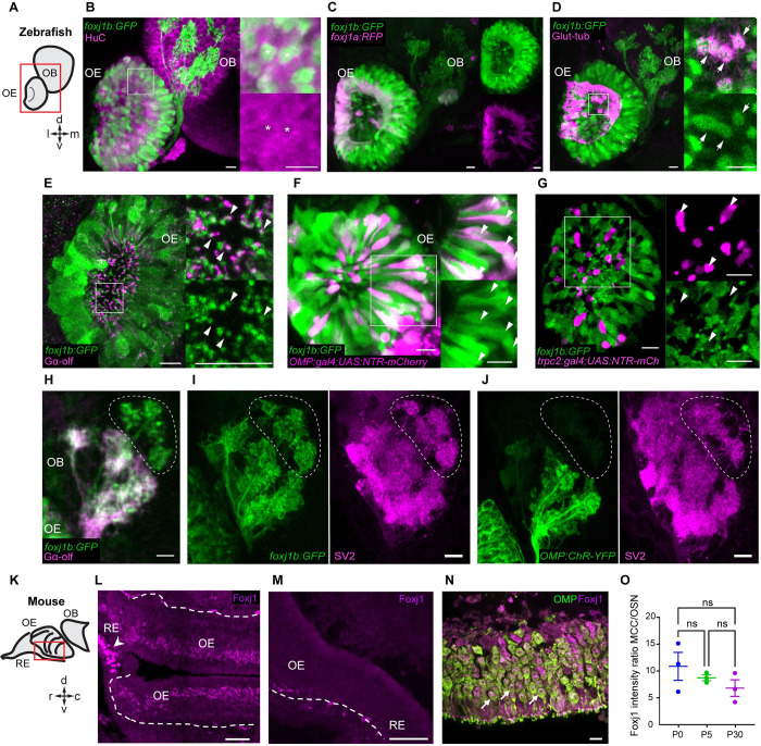
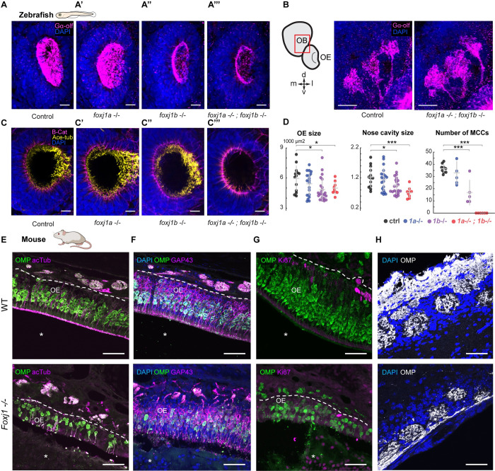
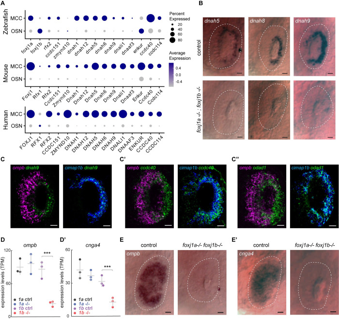
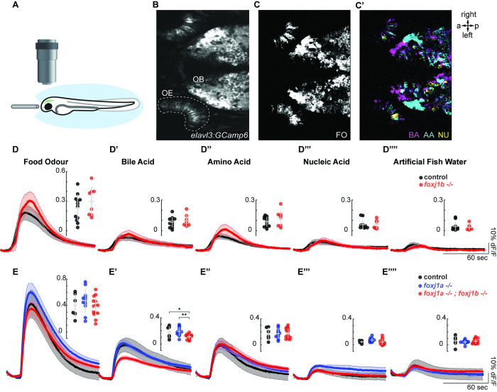
In vertebrates, olfactory receptors localize on multiple cilia elaborated on dendritic knobs of olfactory sensory neurons (OSNs). Although olfactory cilia dysfunction can cause anosmia, how their differentiation is programmed at the transcriptional level has remained largely unexplored. We discovered in zebrafish and mice that Foxj1, a forkhead domain-containing transcription factor traditionally linked with motile cilia biogenesis, is expressed in OSNs and required for olfactory epithelium (OE) formation. In keeping with the immotile nature of olfactory cilia, we observed that ciliary motility genes are repressed in zebrafish, mouse, and human OSNs. Strikingly, we also found that besides ciliogenesis, Foxj1 controls the differentiation of the OSNs themselves by regulating their cell type-specific gene expression, such as that of olfactory marker protein (omp) involved in odor-evoked signal transduction. In line with this, response to bile acids, odors detected by OMP-positive OSNs, was significantly diminished in foxj1 mutant zebrafish. Taken together, our findings establish how the canonical Foxj1-mediated motile ciliogenic transcriptional program has been repurposed for the biogenesis of immotile olfactory cilia, as well as for the development of the OSNs.


Conflict of interest statement

The authors have declared that no competing interests exist.

Figures

Fig 1
Fig 1. foxj1 is expressed in ciliated OSNs of zebrafish and mice.
(A) Schematic showing the larval zebrafish OE and OB; l: lateral, m: medial, d: dorsal, v: ventral. (B-J) Confocal images of the larval (3–5 dpf) zebrafish nose and OB. (B) foxj1b (Gt(foxj1b:GFP), green) is expressed in neurons labeled by HuC (magenta). Asterisks show foxj1-expressing neurons in 5 dpf larva. (C) Differential expression of foxj1a (Gt(foxj1a:2A-TagRFP), magenta) and foxj1b (Gt(foxj1b:GFP), green) paralogs in 5 dpf larvae. Note that foxj1a is expressed primarily at the outer rim of the OE. (D) foxj1b-positive cells (Gt(foxj1b:GFP), green) bear a brush of motile cilia marked by glutamylated-tubulin (magenta) in 5 dpf larvae. (E) foxj1b-positive OSNs (Gt(foxj1b:GFP), green) in the nasal pit bear cilia, indicated by the marker Gαolf (magenta) in 3 dpf larva. White arrowheads in insets show Gαolf marked cilia arising from foxj1b-positive OSNs. (F) Overlap of foxj1b-expressing cells (Gt(foxj1b:GFP), green) with the ciliated OSN marker OMP (Tg(OMP:gal4); Tg(UAS:NTR-mCherry), magenta) in 4 dpf larva. (G) Mutually exclusive localization of foxj1b-expressing cells (Gt(foxj1b:GFP), green) and microvilli OSNs in 4 dpf larvae, indicated by the marker trpc2 (Tg(trpc2:gal4); Tg(UAS:NTR-mCherry), magenta). (H) foxj1b-expressing OSNs (Gt(foxj1b:GFP), green) project mainly to Gαolf-labeled glomeruli (magenta) in 3 dpf larvae. Note that foxj1b:GFP and Gαolf do not overlap entirely (dotted region). (I, J) foxj1b-positive OSNs, in 4 dpf larvae, (I) project to more glomeruli than OMP-expressing OSNs (J) (Tg(OMP:ChR-YFP)). Glomeruli are shown by staining with the presynaptic vesicle marker SV2 (magenta). (K) Schematic showing mouse RE, OE, and OB; r: rostral, c: caudal, d: dorsal, v: ventral. (L) In the nasal epithelium of the adult mouse (P30), Foxj1 localizes to MCCs (arrowhead) within the RE and the layer of OSNs in the OE. Dashed line demarcates the lamina propria separating OE from the underlying tissue. (M) Foxj1 is absent from the nasal epithelium of the Foxj1 knockout mouse (P21). The residual puncta likely represent nonspecific staining of blood vessels or mesenchymal tissue located in the lamina propria under the basement membrane. (N) Foxj1 (magenta) localizes to the nuclei of mature OSNs immunostained for the OMP (P30). (O) Expression levels of Foxj1 in OSNs at different ages (newborn P0, day 5 P5, and adult P30) calculated by comparing immunofluorescence in OSNs relative to respiratory MCCs. No significant difference was found (MCC/OSN ratio of 10.87 ± 2.61, P0; 8.74 ± 0.53, P5; 6.83 ± 1.56, P30; fluorescence intensity was measured in 20–30 cells of each type per individual field of view and ratio was calculated per animal, 3 mice in each group) by one-way ANOVA with Kruskal–Wallis test. Scale bars = 10 μm (B-J), 50 μm (L, M), and 10 μm (N). Raw data files are available in Mendeley Data (https://data.mendeley.com/datasets/2pn963jn6y). dpf, days post fertilization; MCC, motile multiciliated cell; OB, olfactory bulb; OE, olfactory epithelium; OMP, olfactory marker protein; OSN, olfactory sensory neuron; RE, respiratory epithelium.
Fig 2
Fig 2. Foxj1 regulates olfactory cilia biogenesis in zebrafish and OE establishment in mice.
(A) Immunostaining with anti-Gαolf antibody marking olfactory cilia (magenta) in foxj1a,b mutant zebrafish larvae at 4 dpf. Nuclei marked with DAPI (blue). foxj1a mutants showed no observable effect on the formation of olfactory cilia (n = 3). foxj1b mutants showed reduced olfactory cilia (n = 3). foxj1a/b double mutants showed severe loss of olfactory cilia (n = 3). (B, B’) Innervation pattern of ciliated OSNs labeled with Gαolf (magenta) in control (n = 3) and foxj1a/b double mutant (n = 5) showed no significant effect in the formation of olfactory glomeruli at 4 dpf. Nuclei marked with DAPI (blue). (C) foxj1a/b double mutant showing severe reduction of motile cilia number in comparison to the foxj1a mutants, foxj1b mutants and control, as shown by immunostaining with anti-acetylated-tubulin for marking cilia (yellow) and beta-catenin for marking cell borders (magenta) at 4 dpf. (nControl = 13, nfoxj1a−/− = 20, nfoxj1b−/− = 22, nfoxj1a−/−; foxj1b−/− = 9). Nuclei are marked with DAPI (blue). (D) Significant decrease in the size of nose and nasal cavity, and number of MCCs in foxj1b and foxj1a/b double mutant embryos. (For nose size, nControl = 13, nfoxj1a−/− = 20, nfoxj1b−/− = 22, nfoxj1a−/−; foxj1b−/− = 9. For number of MCCs, nControl = 9, nfoxj1a−/− = 5, nfoxj1b−/− = 5, nfoxj1a−/−; foxj1b−/− = 6). (E) The OE of P21 mouse was stained for OMP (green), a mature OSN marker. Apical layer, composed of mucus and cilia, is strongly labeled for acetylated α-tubulin (magenta) in control (top, arrow). Mature OSNs (OMP, green) at the same animal age of 21 day were fewer in number and disorganized within the OE of the Foxj1−/− mouse (bottom), (10,630 ± 923 cells per mm2, n = 8, WT; 3,106 ± 714, n = 7, KO, 3 mice, p = 0.0006). Compared to the WT, thickness of the OE was significantly reduced in the Foxj1−/− mutant (79.68 ± 4.31 μm, n = 34, 3 mice, WT; 45.48 ± 2.15 μm, n = 30, 3 mice, p < 0.0001, KO). (F) Immature OSNs (GAP43, magenta) were located below the layer of mature OSNs (OMP, green) as shown in a control mouse (top). Immature OSNs (GAP43, magenta) lost their orientation relative to mature OSNs and were fewer in numbers within the OE of the Foxj1−/− mutant (bottom) as compared to control (6,339 ± 1,015 cells per mm2, n = 8, WT; 2,008 ± 481, n = 7, KO, 3 mice, p = 0.0012). The nasal cavity (E-G, asterisks) was unobstructed in the WT, whereas in the Foxj1−/− animals, it was completely filled with DAPI and Ki67-positive cells. (G) Proliferating cells expressing Ki67 (magenta) were mostly comprised of basal progenitor cells lining lamina propria. Fewer proliferating cells (Ki67, magenta) were present in the OE of the Foxj1−/− mouse (bottom) as compared to the control (top) (2,362 ± 321 cells per mm2, n = 9, WT; 502 ± 72, n = 9, KO, 3 mice, p < 0.0001). (H) Images showing the OB having oval-shaped glomeruli (top) filled with the axonal projections of OSNs (OMP, white) in control (top). Reduced intensity of OMP immunostaining in the Foxj1−/− mouse (bottom) as compared to the control (top) (352.5 ± 26.7 a.u., n = 21, WT; 266.6 ± 17.5 a.u., n = 44, KO; 3 mice, p = 0.0133). In the Foxj1−/− mouse, glomeruli overall were less developed and had smaller perimeter than in the WT (2,558 ± 368 μm, n = 21, 2 mice, WT; 1,331 ± 306 μm, n = 44, 2 mice, KO; 3 mice, p < 0.0001). Scale bars: 10 μm (A-D), 50 μm (E-H). Raw data files are available in Mendeley Data (https://data.mendeley.com/datasets/2pn963jn6y). dpf, days post fertilization; KO, knockout; MCC, motile multiciliated cell; OB, olfactory bulb; OE, olfactory epithelium; OMP, olfactory marker protein; OSN, olfactory sensory neuron; WT, wild type.
Fig 3
Fig 3. Foxj1 controls the expression of OSN-specific genes, but not ciliary motility genes.
(A) Single-cell transcriptome analysis of ciliated OSNs from wild-type zebrafish, mouse, and human showing low expression of genes encoding axonemal dynein and motility-associated components in OSNs as compared to MCCs. (B) Whole-mount in situ hybridization of dnah5, dnah8, and dnah9 in foxj1a,b double mutant zebrafish embryos at 3 dpf showing absence of gene expression in the periphery of the nasal placodes (highlighted with white dashed line) that primarily consists of OSNs, whereas expression in MCCs at the rim of the cavity (n = 10 for each gene). MCCs are depleted in foxj1a/b double mutants, and, therefore, expression of these genes were absent in the nasal placodes (bottom panel) (n = 5 for dnah5, n = 6 for dnah8, n = 4 for dnah9). Scale bars = 10 μm. (C-C”) Double fluorescent labeling of dnah9 (C), ccdc40 (C’), and odad1 (C”) (green) with ciliated-OSN marker ompb (magenta) and MCC marker cimap1b (blue) by HCR in situ hybridization in wild-type embryo at 3 dpf showing coexpression of dnah9, ccdc40, and odad1 with cimap1b (n = 3), but not with ompb (n = 3). (D, D’) RNA sequencing of foxj1a and foxj1b mutant embryos showed a significant decrease in the expression of the ciliated-OSN marker ompb (D) and cnga4 (D’) in foxj1b mutant embryos but not in foxj1a mutants. (E, F) In situ hybridization in 4 dpf larvae showed reduced expression of ompb (E) and cnga4 (F) in nasal placodes of foxj1a/b double mutant embryos (n = 3). Scale Bars = 20 μm (A-C”), 10 μm (E, E’). Raw data files are available in Mendeley Data (https://data.mendeley.com/datasets/2pn963jn6y). dpf, days post fertilization; HCR, hybridization chain reaction; MCC, motile multiciliated cell; OSN, olfactory sensory neuron.
Fig 4
Fig 4. Olfactory response to bile acid is reduced in foxj1 mutant zebrafish larvae.
(A) Schematic of the olfactory experiment, showing a 4-dpf zebrafish larva embedded in agarose. Its nose was exposed, and odor stimuli were delivered by a fine tube. (B) Neural activity was measured using the Ca2+-reporter GCaMP6s (Tg(elavl3:Gcamp6s)) in a region of interest spanning the entire OE. (C, C’) Representative example showing neural activity in the OE and OB to various odor types. Note that responses in the OB were spatially organized, with nonoverlapping domains. (D-D””) Averaged traces of neural activity on the OE for foxj1b control (n = 12) and mutant (n = 8) fish for each odor type showed no difference in odor responses. Maximum amplitude is shown in the insets. (E-E””) Averaged traces of neural activity in the OE for control (n = 7), foxj1a mutant (n = 10), and double foxj1a/b mutant (n = 14) showed a significant reduction in bile acid response. Shaded error bars are standard error of the mean. Maximum amplitude and standard deviation are shown in the insets. Significance identified by two-sample t test, *: p < 0.05, **: p < 0.01. Raw data files and codes for analysis are available in Mendeley Data (https://data.mendeley.com/datasets/2pn963jn6y). a, anterior; AA, amino acid; BA, bile acid; dpf, days post fertilization; FO, food odor; NU, nucleic acid; OB, olfactory bulb; OE, olfactory epithelium; p, posterior.

Similar articles

Cited by

References

    1. Menco BP, Farbman AI. Genesis of cilia and microvilli of rat nasal epithelia during pre-natal development. II. Olfactory epithelium, a morphometric analysis. J Cell Sci. 1985;78:311–336. doi: 10.1242/jcs.78.1.311 - DOI - PubMed
    1. Ache BW, Young JM. Olfaction: Diverse Species, Conserved Principles. Neuron. 2005;48(3):417–430. - PubMed
    1. McClintock TS, Khan N, Xie C, Martens JR. Maturation of the Olfactory Sensory Neuron and Its Cilia. Chem Senses. 2020;45(9):805–822. doi: 10.1093/chemse/bjaa070 - DOI - PMC - PubMed
    1. Jenkins PM, McEwen DP, Martens JR. Olfactory cilia: linking sensory cilia function and human disease. Chem Senses. 2009;34(5):451–464. doi: 10.1093/chemse/bjp020 - DOI - PMC - PubMed
    1. Kulaga HM, Leitch CC, Eichers ER, Badano JL, Lesemann A, Hoskins BE, et al. Loss of BBS proteins causes anosmia in humans and defects in olfactory cilia structure and function in the mouse. Nat Genet. 2004;36(9):994–998. doi: 10.1038/ng1418 - DOI - PubMed

Substances