. 2024 Jan 25;20(1):e1011983.
doi: 10.1371/journal.ppat.1011983. eCollection 2024 Jan.

BHLHE40 drives protective polyfunctional CD4 T cell differentiation in the female reproductive tract against Chlamydia

Affiliations

BHLHE40 drives protective polyfunctional CD4 T cell differentiation in the female reproductive tract against Chlamydia

Miguel A B Mercado et al. PLoS Pathog. .

Abstract

The protein basic helix-loop-helix family member e40 (BHLHE40) is a transcription factor recently emerged as a key regulator of host immunity to infections, autoimmune diseases and cancer. In this study, we investigated the role of Bhlhe40 in protective T cell responses to the intracellular bacterium Chlamydia in the female reproductive tract (FRT). Mice deficient in Bhlhe40 exhibited severe defects in their ability to control Chlamydia muridarum shedding from the FRT. The heightened bacterial burdens in Bhlhe40-/- mice correlated with a marked increase in IL-10-producing T regulatory type 1 (Tr1) cells and decreased polyfunctional CD4 T cells co-producing IFN-γ, IL-17A and GM-CSF. Genetic ablation of IL-10 or functional blockade of IL-10R increased CD4 T cell polyfunctionality and partially rescued the defects in bacterial control in Bhlhe40-/- mice. Using single-cell RNA sequencing coupled with TCR profiling, we detected a significant enrichment of stem-like T cell signatures in Bhlhe40-deficient CD4 T cells, whereas WT CD4 T cells were further down on the differentiation trajectory with distinct effector functions beyond IFN-γ production by Th1 cells. Altogether, we identified Bhlhe40 as a key molecular driver of CD4 T cell differentiation and polyfunctional responses in the FRT against Chlamydia.


Conflict of interest statement

The authors have declared that no competing interests exist.

Figures
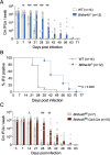
Fig 1
Fig 1. Bhlhe40-/- and Bhlhe40fl/fl-Cd4-Cre mice exhibit delayed bacterial clearance following Chlamydia muridarum intravaginal infection.
(A-B) WT and Bhlhe40-/- mice were infected intravaginally with 1×10⁵ C. muridarum. Bacterial shedding from the lower female reproductive tract (FRT) (A) and percentage of bacterial clearance (B) were monitored by vaginal swabs. Data are combined results of three independent experiments with 12 to 14 mice per group. (C) Bhlhe40fl/fl and Bhlhe40fl/fl-Cd4-Cre mice were infected intravaginally with 1×10⁵ C. muridarum. Bacterial shedding was monitored by vaginal swabs. Data are combined results of two independent experiments with 7 to 10 mice per group. Each data point represents an individual mouse. Error bars represent the mean ± SEM. ****p < 0.0001 by two-way ANOVA and **p < 0.01, ***p < 0.001 by ANOVA multiple comparisons in (A) and (C); ****p < 0.0001 by Log-rank in (B).
Fig 2
Fig 2. Bhlhe40 suppresses Chlamydia-specific Tr1 differentiation in a T cell-intrinsic manner.
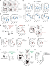
(A-C) WT and Bhlhe40-/- mice were infected intravaginally with 1×10⁵ C. muridarum and analyzed at 10 dpi. (A) Representative FACS plots depicting IFNγ- and IL-10-producing CD4 T cells (gated on live CD90.2+CD4+CD44hi cells). (B) Percentages and total cell numbers of IFNγ+, IL-10+ and IFNγ+IL-10+ CD4 T cells within the CD4+CD44hi population. (C) Mean fluorescence intensities (MFIs) of IFNγ and IL-10 in CD4+CD44hiIFNγ+ and CD4+CD44hiIL-10+ T cells, respectively. Data are from two independent experiments with 8 mice per group. Each data point represents an individual mouse. (D-H) Mixed WT and Bhlhe40-/- CD4 T cell adoptive transfer. (D) Experimental workflow. (E) Normalized ratios of Bhlhe40-/- (CD45.2+) and WT (CD45.1+) donor CD4 T cells in TCRβ-/- host. Representative FACS plots (F), summary data (G) and MFI (H) of cytokine-producing donor CD4 T cells in TCRβ-/- FRT. Data are from two independent experiments with 6 mice per group. Each pair of data points represents the two donor T cell populations in an individual TCRβ-/- host. (I-K) Chlamydia-specific TP1 CD4 T cell adoptive transfer. (I) Experimental workflow. Representative FACS plots (J) and summary data (K) of cytokine-producing Bhlhe40-/- TP1 (CD45.2+CD90.1-), WT TP1 (CD45.2+CD90.1+) and B6 host (CD45.2-CD90.1-) CD4 T cells in the host FRT. Data are from two independent experiments with 6 samples per group. Each data point represents a pooled FRT sample from 4 mice. Error bars represent the mean ± SEM. *p < 0.05, **p < 0.01, ***p < 0.001, ns, not significant by unpaired t test in (B-C), and paired t test in (E-K).
Fig 3
Fig 3. Anti-IL-10R blockade partially restores the ability of bacterial control in Bhlhe40-deficient mice.
WT, Bhlhe40-/- and Il10-/- mice were infected intravaginally with 1×10⁵ C. muridarum. Cohorts of WT and Bhlhe40-/- mice were treated with anti-IL-10R or isotype control antibody twice a week until day 21. Bacterial shedding from the lower FRT were monitored by vaginal swabs. Data are results from two independent experiments with 7–8 mice per group. Each data point represents an individual mouse. Error bars represent the mean ± SEM. ****p < 0.0001 by one-way ANOVA and *p < 0.05, **p < 0.01, ***p < 0.001 by ANOVA multiple comparisons.
Fig 4
Fig 4. CD4 T cell polyfunctionality inversely correlates with host susceptibility to C. muridarum in the FRT.
(A) WT and Bhlhe40-/- mice were infected intravaginally with 1×10⁵ C. muridarum. Cytokine levels in the supernatants of ex vivo cultures of DLN and FRT cells as measured at 10 dpi. Data are from two independent experiments with 9 mice per group. Each data point represents an individual mouse. (B-D) WT, Bhlhe40-/-, Bhlhe40-/- x Il10-/- and Il10-/- mice were infected intravaginally with 1×10⁵ C. muridarum. Representative FACS plots (B), SPICE analysis (C) and summary data (D) depicting cytokine producing CD4 T cells (gated on live CD90.2+CD4+CD44hiIL-10- cells) at 14 dpi. Data are from two independent experiments with 6–9 mice per group. Each data point represents an individual mouse. (E-F) Chlamydia-specific TP1 CD4 T cell adoptive transfer experiment (see workflow in Fig 2I). Representative FACS plots (E) and summary data (F) of cytokine-producing Bhlhe40-/- TP1 (CD45.2+CD90.1-), WT TP1 (CD45.2+CD90.1+) and B6 host CD4 T cells (CD45.2-CD90.1-) in the host FRT. Data are from two independent experiments with 6 samples per group. Each data point represents a pooled FRT sample from 4 mice. (G) Polyfunctionality Index (left) and bacterial burdens (right) in WT, Bhlhe40-/-, Bhlhe40-/- x Il10-/- and Il10-/- mice. Data are from two independent experiments with 6–9 mice per group. Each data point represents an individual mouse. Error bars represent the mean ± SEM. *p < 0.05, **p < 0.01, ***p < 0.001 by unpaired t test in (A), (D) and (G), and paired t test in (F).
Fig 5
Fig 5. Single-cell RNA sequencing detects stem-like CD4 T cells in Bhlhe40-/- FRT.
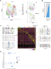
WT and Bhlhe40-/- mice were infected intravaginally with 1×10⁵ C. muridarum. Activated CD4 T cells (live CD90.2+CD4+CD44hi) in the FRT were sorted for scRNAseq using 10x genomics 5’ GEX. (A) UMAP depicting 14 clusters of CD4 T cells from merged WT and Bhlhe40-/- CD4 T cells (left); deconvolution of the composite UMAP into WT and Bhlhe40-/- components. (B) Percentages of WT and Bhlhe40-/- CD4 T cells in each cluster following normalization of the two populations into the same cell numbers. (C) Heatmap showing top 30 most upregulated genes in each cluster, and representative genes specifically expressed within each of the top 3 clusters in WT and Bhlhe40-/- CD4 T cells are shown on the sides with violin plots depicting expression levels in each cluster. (D) Dot plots depicting Th lineage-specific cytokines and transcription factors.
Fig 6
Fig 6. Pseudotime trajectory and TCR clonal type analyses reveal CD4 T cell differentiation and clonal expansion profiles in WT and Bhlhe40-/- mice.
(A) Pseudotime trajectory analysis of CD4 T cell clusters in Fig 5 as conducted using Monocle 3. (B) WT and Bhlhe40-/- CD4 T cells were binned into quartiles based on clonotype abundance. Shaded area represents the top 25% (expanders). (C) UMAP showing the expanders in WT and Bhlhe40-/- CD4 T cell clusters. (D) Frequencies of expanders in each cluster in WT and Bhlhe40-/- CD4 T cells.

Update of

References

    1. Zhu J, Yamane H, Paul WE. Differentiation of Effector CD4 T Cell Populations. Annu Rev Immunol. 2010;28: 445–489. doi: 10.1146/annurev-immunol-030409-101212 - DOI - PMC - PubMed
    1. Helble J D., Starnbach M N. T cell responses to Chlamydia. Pathogens and Disease. 2021;79: ftab014. doi: 10.1093/femspd/ftab014 - DOI - PMC - PubMed
    1. Perry LL, Feilzer K, Caldwell HD. Immunity to Chlamydia trachomatis is mediated by T helper 1 cells through IFN-gamma-dependent and -independent pathways. The Journal of Immunology. 1997;158: 3344–3352. Available: https://www.jimmunol.org/content/158/7/3344 - PubMed
    1. Coers J, Gondek DC, Olive AJ, Rohlfing A, Taylor GA, Starnbach MN. Compensatory T Cell Responses in IRG-Deficient Mice Prevent Sustained Chlamydia trachomatis Infections. PLOS Pathogens. 2011;7: e1001346. doi: 10.1371/journal.ppat.1001346 - DOI - PMC - PubMed
    1. Haldar AK, Piro AS, Finethy R, Espenschied ST, Brown HE, Giebel AM, et al.. Chlamydia trachomatis Is Resistant to Inclusion Ubiquitination and Associated Host Defense in Gamma Interferon-Primed Human Epithelial Cells. mBio. 2016;7: e01417–16. doi: 10.1128/mBio.01417-16 - DOI - PMC - PubMed

MeSH terms

Substances