

Photon Counting Computed Tomography with the Radiation Dose of a Chest X-Ray: Feasibility and Diagnostic Yield
- PMID: 38272004
- PMCID: PMC10871675
- DOI: 10.1159/000536065
Photon Counting Computed Tomography with the Radiation Dose of a Chest X-Ray: Feasibility and Diagnostic Yield
Abstract
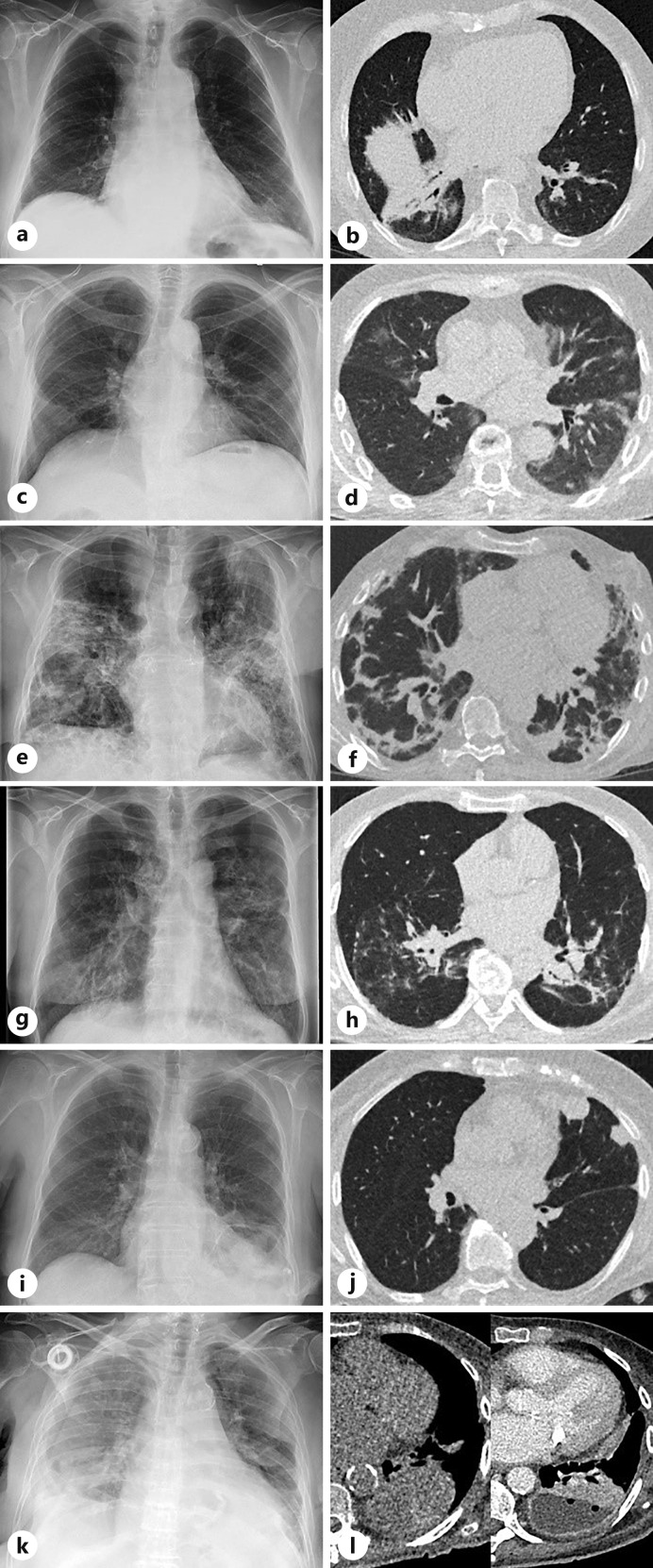
Introduction: Photon counting (PC) detectors allow a reduction of the radiation dose in CT. Chest X-ray (CXR) is known to have a low sensitivity and specificity for detection of pneumonic infiltrates. The aims were to establish an ultra-low-dose CT (ULD-CT) protocol at a PC-CT with the radiation dose comparable to the dose of a CXR and to evaluate its clinical yield in patients with suspicion of pneumonia.
Methods: A ULD-CT protocol was established with the aim to meet the radiation dose of a CXR. In this retrospective study, all adult patients who received a ULD-CT of the chest with suspected pneumonia were included. Radiation exposure of ULD-CT and CXR was calculated. The clinical significance (new diagnosis, change of therapy, additional findings) and limitations were evaluated by a radiologist and a pulmonologist considering previous CXR and clinical data.
Results: Twenty-seven patients (70% male, mean age 68 years) were included. With our ULD-CT protocol, the radiation dose of a CXR could be reached (mean radiation exposure 0.11 mSv). With ULD-CT, the diagnosis changed in 11 patients (41%), there were relevant additional findings in 4 patients (15%), an infiltrate (particularly fungal infiltrate under immunosuppression) could be ruled out with certainty in 10 patients (37%), and the therapy changed in 10 patients (37%). Two patients required an additional CT with contrast medium to rule out a pulmonary embolism or pleural empyema.
Conclusions: With ULD-CT, the radiation dose of a CXR could be reached while the clinical impact is higher with change in diagnosis in 41%.
Keywords: Community-acquired pneumonia; Photon counting computed tomography; Ultra-low-dose computed tomography.
© 2024 The Author(s). Published by S. Karger AG, Basel.
Conflict of interest statement
The authors of this manuscript declare relationships with the following companies: S.D. reports grants from Siemens Healthineers paid to the institution and personal honoraria for lectures from !DE Werbeagentur GmbH, Boehringer Ingelheim, and med update GmbH. T.W., V.N.M., T.B., O.J., J.V.-C., and T.W. declare that they have no competing interests. F.W. reports grants unrelated to this publication from the German Ministry of Research and Education (BMBF), German Cancer Aid, Siemens Healthineers, Promedicus, and Delcath paid to his institution. J.R. reports grants from the German Center for Lung Research (DZL), the German Center for Infection Research (DZIF), the Bundesministerium für Bildung und Forschung (BMBF), and the Bundesministerium für Gesundheit (BMG) paid to her institution; and personal honoraria for lectures from AstraZeneca, Berlin Chemie, Insmed, GSK, MSD, Shionogi, and Thermo Fisher personal payments for participation on an advisory board from Insmed, GSK, GILEAD, MSD, Thermo Fisher, and Shionogi.
Figures
Similar articles
-
Diagnostic accuracy of ultra-low-dose chest computed tomography in an emergency department.Acta Radiol. 2022 Mar;63(3):336-344. doi: 10.1177/0284185121995804. Epub 2021 Mar 4. Acta Radiol. 2022. PMID: 33663246
-
Ultra-low-dose computed tomography and chest X-ray in follow-up of high-grade soft tissue sarcoma-a prospective comparative study.Sci Rep. 2024 Mar 26;14(1):7181. doi: 10.1038/s41598-024-57770-z. Sci Rep. 2024. PMID: 38531939 Free PMC article.
-
Ultra-low dose chest CT for the diagnosis of pulmonary arteriovenous malformation in patients with hereditary hemorrhagic telangiectasia.Diagn Interv Imaging. 2024 Oct;105(10):364-370. doi: 10.1016/j.diii.2024.03.006. Epub 2024 Apr 10. Diagn Interv Imaging. 2024. PMID: 38604894
-
Diagnostic Accuracy of Low and Ultra-Low Dose CT for Identification of Urinary Tract Stones: A Systematic Review.Urol Int. 2018;100(4):375-385. doi: 10.1159/000488062. Epub 2018 Apr 12. Urol Int. 2018. PMID: 29649823
-
[What future for chest x-ray against ultra-low-dose computed tomography?].Rev Pneumol Clin. 2017 Feb;73(1):3-12. doi: 10.1016/j.pneumo.2016.09.007. Epub 2016 Dec 9. Rev Pneumol Clin. 2017. PMID: 27956084 Review. French.
Cited by
-
Imaging the Lung in ARDS: A Primer.Respir Care. 2024 Jul 24;69(8):1011-1024. doi: 10.4187/respcare.12061. Respir Care. 2024. PMID: 39048146 Free PMC article. Review.
-
First assessment of photon-counting CT for virtual bronchoscopic navigation.Eur Respir J. 2025 Apr 3;65(4):2402476. doi: 10.1183/13993003.02476-2024. Print 2025 Apr. Eur Respir J. 2025. PMID: 39915055 Free PMC article.
-
Paediatric high-pitch lung imaging with photon-counting detector computed tomography: a dose reduction phantom study.Pediatr Radiol. 2025 May;55(6):1191-1201. doi: 10.1007/s00247-025-06235-0. Epub 2025 Apr 15. Pediatr Radiol. 2025. PMID: 40229451 Free PMC article.
-
Photon-Counting CT in Cancer Radiotherapy: Technological Advances and Clinical Benefits.ArXiv [Preprint]. 2024 Dec 4:arXiv:2410.20236v3. ArXiv. 2024. Update in: Phys Med Biol. 2025 May 16;70(10). doi: 10.1088/1361-6560/add4ba. PMID: 39575116 Free PMC article. Updated. Preprint.
-
Photon-counting CT in cancer radiotherapy: technological advances and clinical benefits.Phys Med Biol. 2025 May 16;70(10):10TR01. doi: 10.1088/1361-6560/add4ba. Phys Med Biol. 2025. PMID: 40328288 Free PMC article. Review.
References
-
- van der Bie J, van Straten M, Booij R, Bos D, Dijkshoorn ML, Hirsch A, et al. . Photon-counting CT: review of initial clinical results. Eur J Radiol. 2023;163:110829. - PubMed
-
- Flohr T, Petersilka M, Henning A, Ulzheimer S, Ferda J, Schmidt B. Photon-counting CT review. Phys Med. 2020;79:126–36. - PubMed
-
- Willemink MJ, Persson M, Pourmorteza A, Pelc NJ, Fleischmann D. Photon-counting CT: technical principles and clinical prospects. Radiology. 2018;289(2):293–312. - PubMed
