. 2024 Feb 1;111(2):364-382.
doi: 10.1016/j.ajhg.2023.12.016. Epub 2024 Jan 24.

Role of CAMK2D in neurodevelopment and associated conditions

Affiliations

Role of CAMK2D in neurodevelopment and associated conditions

Pomme M F Rigter et al. Am J Hum Genet. .

Erratum in

  • Role of CAMK2D in neurodevelopment and associated conditions.
    Rigter PMF, de Konink C, Dunn MJ, Proietti Onori M, Humberson JB, Thomas M, Barnes C, Prada CE, Weaver KN, Ryan TD, Caluseriu O, Conway J, Calamaro E, Fong CT, Wuyts W, Meuwissen M, Hordijk E, Jonkers CN, Anderson L, Yuseinova B, Polonia S, Beysen D, Stark Z, Savva E, Poulton C, McKenzie F, Bhoj E, Bupp CP, Bézieau S, Mercier S, Blevins A, Wentzensen IM, Xia F, Rosenfeld JA, Hsieh TC, Krawitz PM, Elbracht M, Veenma DCM, Schulman H, Stratton MM, Küry S, van Woerden GM. Rigter PMF, et al. Am J Hum Genet. 2025 Sep 4;112(9):2247. doi: 10.1016/j.ajhg.2025.08.001. Epub 2025 Aug 11. Am J Hum Genet. 2025. PMID: 40795868 Free PMC article. No abstract available.

Abstract

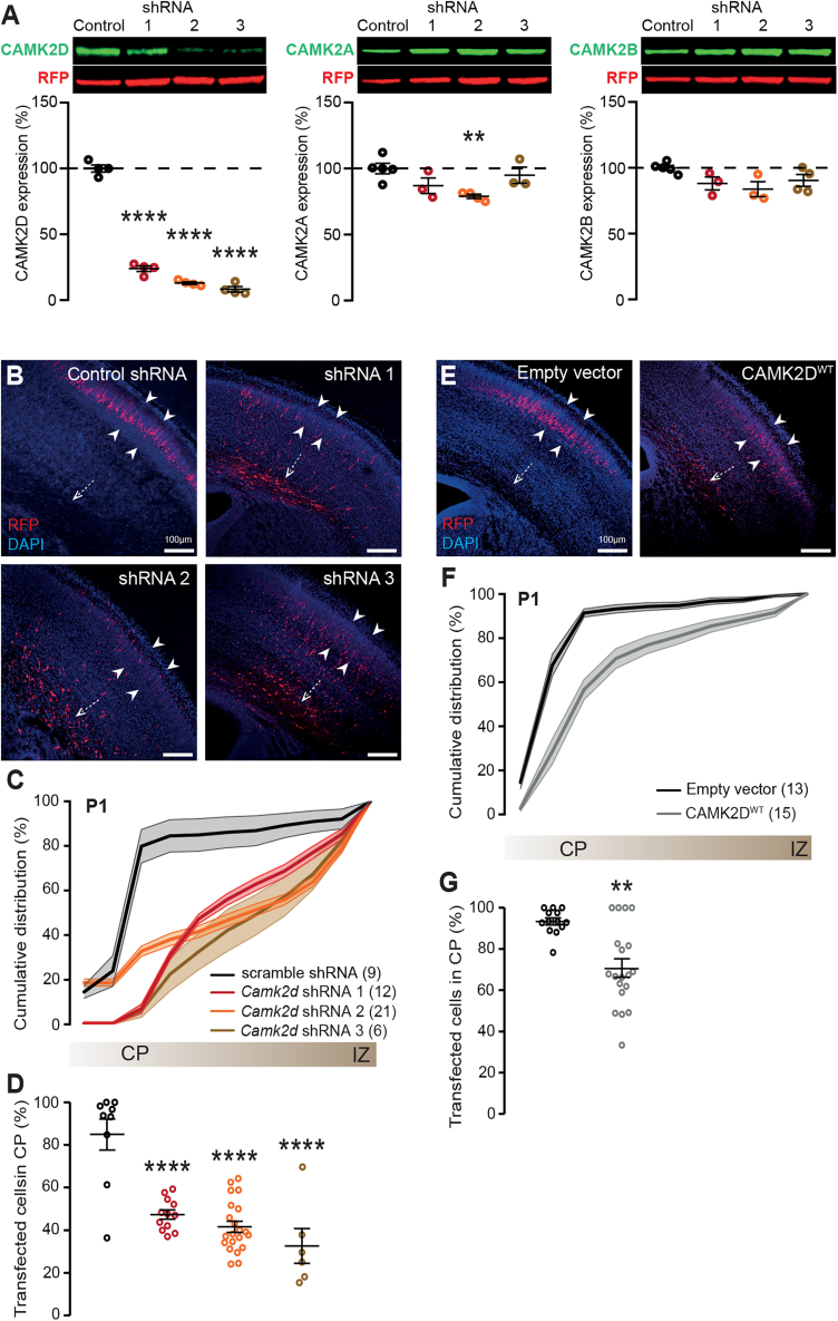
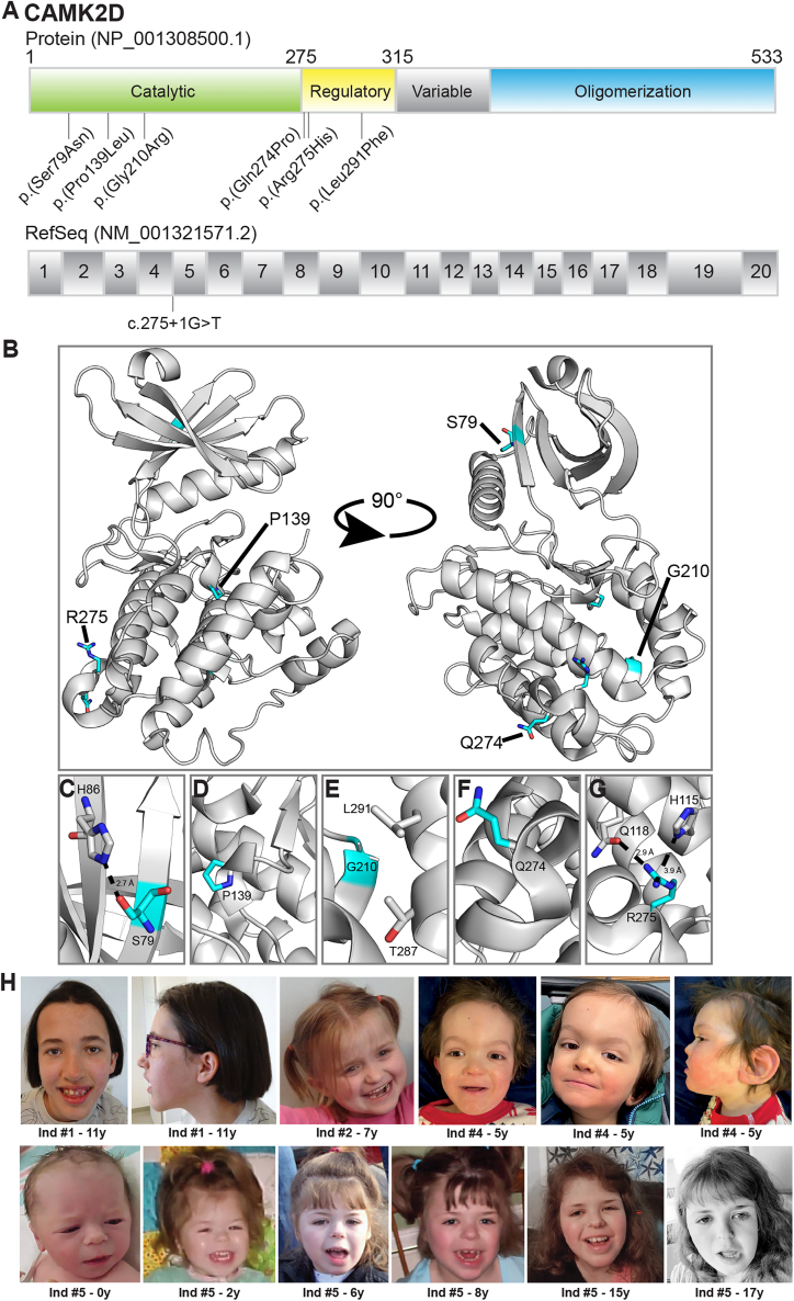
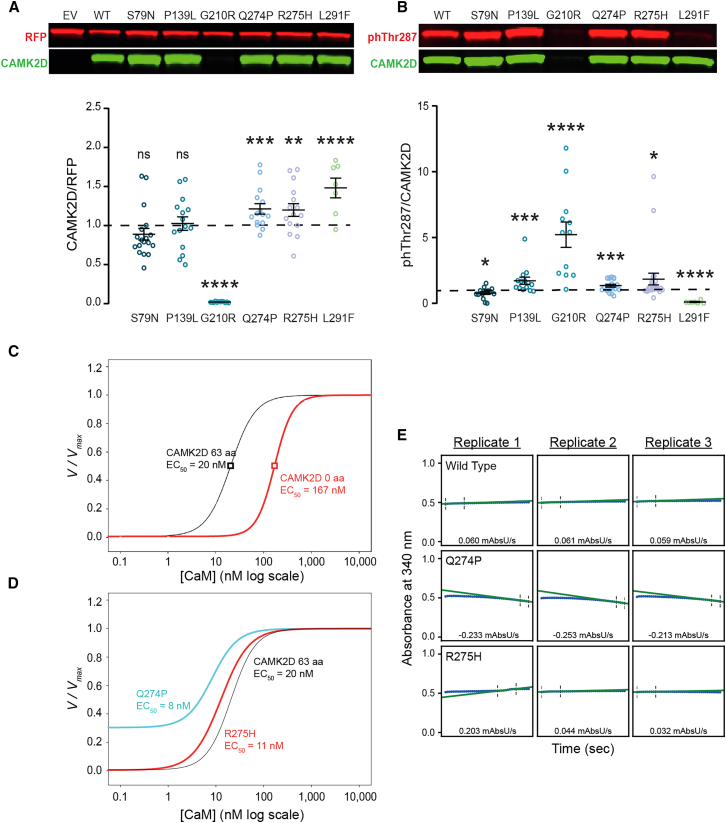
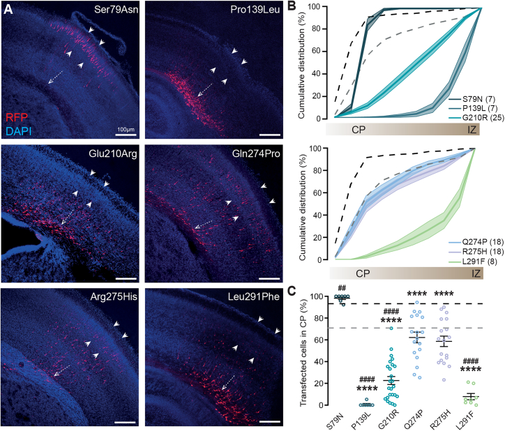
The calcium/calmodulin-dependent protein kinase type 2 (CAMK2) family consists of four different isozymes, encoded by four different genes-CAMK2A, CAMK2B, CAMK2G, and CAMK2D-of which the first three have been associated recently with neurodevelopmental disorders. CAMK2D is one of the major CAMK2 proteins expressed in the heart and has been associated with cardiac anomalies. Although this CAMK2 isoform is also known to be one of the major CAMK2 subtypes expressed during early brain development, it has never been linked with neurodevelopmental disorders until now. Here we show that CAMK2D plays an important role in neurodevelopment not only in mice but also in humans. We identified eight individuals harboring heterozygous variants in CAMK2D who display symptoms of intellectual disability, delayed speech, behavioral problems, and dilated cardiomyopathy. The majority of the variants tested lead to a gain of function (GoF), which appears to cause both neurological problems and dilated cardiomyopathy. In contrast, loss-of-function (LoF) variants appear to induce only neurological symptoms. Together, we describe a cohort of individuals with neurodevelopmental disorders and cardiac anomalies, harboring pathogenic variants in CAMK2D, confirming an important role for the CAMK2D isozyme in both heart and brain function.

Keywords: CAMK2D; calcium/calmodulin-dependent protein kinase 2 delta; cardiomyopathy; intellectual disability; neurodevelopment.


Conflict of interest statement

Declaration of interests The Department of Molecular and Human Genetics at Baylor College of Medicine receives revenue from clinical genetic testing completed at Baylor Genetics Laboratories. A.B. and I.M.W. are employees of GeneDx, LLC; H.S. is a consultant for Vasa Therapeutics (Poland).

Figures

Figure 1
Figure 1
Reduced as well as increased CAMK2D protein level during prenatal neurodevelopment results in a migration delay (A) Knockdown of Camk2d with shRNAs proves to be efficient and mostly specific. Top: representative western blots of protein lysates from HEK293T cells, which were transfected with either control (scramble) or Camk2d specific shRNA’s. Blots were probed with an antibody against the specific CAMK2 (green), and RFP (red). Bottom: Quantification of CAMK2D (left), CAMK2A (middle), or CAMK2B (right) protein levels normalized against RFP. (B) Representative images of coronal brain slices from P0/P1 pups that were transfected with scramble or Camk2d-specific shRNAs at E14.5, using in utero electroporation. White dashed arrow indicates the subventricular zone (SVZ); white arrowheads indicate the cortical plate (CP). DAPI is in blue, RFP in red. (C) Cumulative graph indicating the migration pattern from the SVZ to the CP in presence of scramble or Camk2d-specific shRNAs. (D) Quantification of tdTomato-positive cells that have successfully migrated to the CP, revealing that knockdown of Camk2d leads to a delay in migration. (E) Representative images of coronal brain slices from P0/P1 pups that were transfected with a control empty vector or CAMK2DWT at E14.5, using in utero electroporation. (F) Cumulative graph indicating the migration pattern from the SVZ to the CP upon overexpression of the empty vector or CAMK2DWT. (G) Quantification of tdTomato-positive cells that have successfully migrated to the CP, showing that overexpression of CAMK2DWT leads to a delay in migration as well. Number in parentheses represents the number of images used for the quantification; dots represent data points and error bars indicate SEM; p < 0.05; ∗∗p < 0.01; ∗∗∗∗p < 0.0001.
Figure 2
Figure 2
CAMK2D missense variants identified in our cohort (A) Schematic overview of the CAMK2D protein domain organization (top) and the corresponding messenger CAMK2D RNA structure (bottom), showing the location of the different missense variants as well as the location of the splice site variant. Four of the variants are located in the catalytic domain (c.275+1G>T, p.Ser79Asn, p.Pro139Leu, and p.Gly210Arg) and three are located in the regulatory domain (p.Gln274Pro, p.Arg275His, and p.Leu291Phe). (B) Crystal structure of inhibited CAMK2D and regulatory segment (PDB: 2VN944), with the positions of the human variants highlighted as cyan sticks. (C) The hydroxyl side chain of Ser79 forms an electrostatic interaction with the sidechain of His86. Asn at this position would be too bulky to accommodate this interaction. (D) Pro139 facilitates a short helical turn in the C-lobe. Substitution to Leu would disrupt this. (E) Gly210 is located at the base of a helix directly facing the regulatory segment. Arg at this position would be too bulky and likely dislodge the regulatory segment. (F) Gln274 is located within a helical turn. The positioning of a Pro in this helix would disrupt folding of this region. (G) Arg275 is located within the same turn and is proximal enough to Gln118 for hydrogen bonding (2.9 Å) and His115 (3.9 Å) for like-charged pair interaction between the two nitrogen atoms. Thus, substitution to a His would disrupt these interactions and the position of the alpha helix. (H) Pictures of four individuals carrying a variant in CAMK2D. Note the coarse features and down-slanting palpebral fissures visible especially in individuals #2, #4, and #5, who also have a short nose and thin lips. Individual #1 has a long face with a high forehead, and she also has low-set ears, like individual #4.
Figure 3
Figure 3
Assessment of protein levels and kinase activity (A) Top: representative western blot of protein lysates from HEK293T cells that were transfected with either EV (empty control vector), CAMK2DWT, CAMK2DS79N, CAMK2DP139L, CAMK2DG210R, CAMK2DQ274P, CAMK2DR275H, or CAMK2DL291F, probed with an antibody against CAMK2D (green) and RFP (red). Bottom: Quantification of CAMK2D expression normalized against RFP, showing the effects on protein of the variants. Black dashed line represents CAMK2DWT. (B) Top: representative western blot of protein lysates from HEK293T cells which were transfected with either EV (empty control vector), CAMK2DWT, CAMK2DS79N, CAMK2DP139L, CAMK2DG210R, CAMK2DQ274P, CAMK2DR275H, or CAMK2DL291F, probed with an antibody against CAMK2D (green), phThr287 (red). Bottom: Quantification of autophosphorylated CAMK2D (phThr287) normalized against CAMK2D protein level, showing the effects on autophosphorylation of the variants. All data were normalized against level of autophosphorylation in CAMK2DWT, represented by the black dashed line. (C and D) On the y axis is the normalized rate of phosphorylation and on the x axis the respective calmodulin concentration. Data were normalized within the triplicate to the maximal rate. The hill coefficient (nH) and EC50 determined by the fits are listed below the corresponding CAMK2D variant for each plot. (C) Fits (see subjects and methods) of CAMK2D without linker (red) or with a 63-amino-acid linker region (black). (D) Fits of CAMK2DQ274P (cyan) and CAMK2DR275P (red) each with a 63-amino acid linker. (E) Individual replicates of the coupled kinase assay in absence of CaM, to assess baseline kinase activity. A negative slope indicates increased kinase activity (blue line is the raw data; green line is the linear fit from which maximal slopes are calculated). Dots represent data points. Error bars indicate SEM; p < 0.05; ∗∗p < 0.01; ∗∗∗p < 0.001; ∗∗∗∗p < 0.0001.
Figure 4
Figure 4
Overexpression of CAMK2D variants during prenatal neurodevelopment causes a migration deficit (A) Representative images of coronal brain slices from P0/P1 pups that were transfected with either CAMK2DS79N, CAMK2DP139L, CAMK2DG210R, CAMK2DQ274P, CAMK2DR275H, or CAMK2DL291F at E14.5, using in utero electroporation. White dashed arrow indicates the SVZ; white arrowheads indicate the CP. DAPI in blue, RFP in red. Scale bar indicates 100 μm. (B) Cumulative graph indicating the migration pattern from the SVZ to the CP in presence of overexpressed CAMK2DS79N, CAMK2DP139L, CAMK2DG210R, CAMK2DQ274P, CAMK2DR275H, or CAMK2DL291F. Number in parentheses is the number of images used for the quantification. Black dashed line indicates EV (n = 12); gray dashed line indicates CAMK2DWT (n = 19) overexpression. (C) Quantification of RFP-positive cells that have successfully migrated to the CP, showing the different effects of the variants on migration. # indicates the levels of significance compared to CAMK2DWT overexpression. indicates the levels of significance compared to the empty vector control (EV). Black dashed line represents EV, gray dashed line represents CAMK2DWT overexpression. Dots represent data points. Error bars indicate SEM; ∗∗/##p < 0.01; ∗∗∗∗/####p < 0.0001.

References

    1. Bhattacharyya M., Stratton M.M., Going C.C., McSpadden E.D., Huang Y., Susa A.C., Elleman A., Cao Y.M., Pappireddi N., Burkhardt P., et al. Molecular mechanism of activation-triggered subunit exchange in Ca2+/calmodulin-dependent protein kinase II. Elife. 2016;5 - PMC - PubMed
    1. Lisman J., Yasuda R., Raghavachari S. Mechanisms of CaMKII action in long-term potentiation. Nat. Rev. Neurosci. 2012;13:169–182. - PMC - PubMed
    1. Hell J.W. CaMKII: Claiming center stage in postsynaptic function and organization. Neuron. 2014;81:249–265. - PMC - PubMed
    1. Gaertner T.R., Kolodziej S.J., Wang D., Kobayashi R., Koomen J.M., Stoops J.K., Waxham M.N. Comparative Analyses of the Three-dimensional Structures and Enzymatic Properties of α, β, γ, and δ Isoforms of Ca2+-Calmodulin-dependent Protein Kinase II. J. Biol. Chem. 2004;279:12484–12494. - PubMed
    1. Hudmon A., Schulman H. Neuronal Ca2+/Calmodulin-Dependent Protein Kinase II: The Role of Structure and Autoregulation in Cellular Function. Annu. Rev. Biochem. 2002;71:473–510. - PubMed

Publication types

MeSH terms

Substances