. 2024 Jan;25(1):e12927.
doi: 10.1111/tra.12927.

Rescue of secretion of rare-disease-associated misfolded mutant glycoproteins in UGGT1 knock-out mammalian cells

Affiliations

Rescue of secretion of rare-disease-associated misfolded mutant glycoproteins in UGGT1 knock-out mammalian cells

Gabor Tax et al. Traffic. 2024 Jan.

Abstract

Endoplasmic reticulum (ER) retention of misfolded glycoproteins is mediated by the ER-localized eukaryotic glycoprotein secretion checkpoint, UDP-glucose glycoprotein glucosyl-transferase (UGGT). The enzyme recognizes a misfolded glycoprotein and flags it for ER retention by re-glucosylating one of its N-linked glycans. In the background of a congenital mutation in a secreted glycoprotein gene, UGGT-mediated ER retention can cause rare disease, even if the mutant glycoprotein retains activity ("responsive mutant"). Using confocal laser scanning microscopy, we investigated here the subcellular localization of the human Trop-2-Q118E, E227K and L186P mutants, which cause gelatinous drop-like corneal dystrophy (GDLD). Compared with the wild-type Trop-2, which is correctly localized at the plasma membrane, these Trop-2 mutants are retained in the ER. We studied fluorescent chimeras of the Trop-2 Q118E, E227K and L186P mutants in mammalian cells harboring CRISPR/Cas9-mediated inhibition of the UGGT1 and/or UGGT2 genes. The membrane localization of the Trop-2 Q118E, E227K and L186P mutants was successfully rescued in UGGT1-/- cells. UGGT1 also efficiently reglucosylated Trop-2-Q118E-EYFP in cellula. The study supports the hypothesis that UGGT1 modulation would constitute a novel therapeutic strategy for the treatment of pathological conditions associated to misfolded membrane glycoproteins (whenever the mutation impairs but does not abrogate function), and it encourages the testing of modulators of ER glycoprotein folding quality control as broad-spectrum rescue-of-secretion drugs in rare diseases caused by responsive secreted glycoprotein mutants.

Keywords: GDLD; TACSTD2; Trop-2; UGGT; UGGT1; UGGT2; glycoprotein secretion; responsive mutant.


Conflict of interest statement

Conflicts of Interest: Nil.

Figures

Figure 1.
Figure 1.. Rescue of secretion of the Trop-2-Q118E-EYFP- mutant glycoprotein upon UGGT1 inactivation.
Confocal images of single optical sections of transiently transfected HEK293T (live imaging) and Vero E6 cells (fixed cells).Nuclei were stained with Hoechst 33342 in HEK293T cells or with DAPI stain in Vero E6 cells (blue). (A, B): Wild type (WT) and UGGT1−/−HEK293T and Vero E6 cells were transiently transfected with Trop-2-pEYFP-N1 and Trop-2-Q118E-pEYFP-N1 plasmids. (A): The Trop-2-EYFP wild type glycoprotein (green) traverses the secretory pathway and reaches the plasma membrane (white arrowheads);(B): in WT cells, the Trop-2-Q118E-EYFP mutant glycoprotein (green) is visible in the secretory pathway but absent from the plasma membrane. In the UGGT1−/− cells, the same Trop-2-Q118E-EYFP mutant glycoprotein reaches the cellular membrane (white arrowheads). (C, D) WT, UGGT1−/−, UGGT2−/−, and UGGT1/2−/−HEK293T cells were transiently transfected with Trop-2-pEYFP-N1 (C) and Trop-2-Q118E-pEYFP-N1 plasmids (D). Wheat Germ Agglutinin (WGA) Alexa Fluor 555 (orange) enables verification of plasma membrane localisation of the fluorescent fusion glycoproteins; (C): in both WT and UGGT1/− HEK293T cells the Trop-2-EYFP-WT glycoprotein (green) reaches the cellular membrane (white arrowheads); (D): in the WT HEK293T cells the Trop-2-Q118E-EYFP mutant glycoprotein (green) is trapped in the secretory pathway, while in the UGGT1/−and UGGT1/2−/−HEK293T cells (and to a lower extent in UGGT2−/− cells) the same mutant glycoprotein is visible both in the ER and in the membrane (white arrowheads).
Figure 2.
Figure 2.. Multichannel intensity plots of the Trop-2-pEYFP-N1 transfected HEK293T cell lines.
For each combination of cell line and transiently transfected vector, the fluorescent signal is plotted along the line drawn in white in the inset panel. Labelling: green = EYFP; orange = WGA Alexa Fluor 555; blue = Hoechst 33342. The signal from the WGA Alexa Fluor 555 labelled membrane (orange) overlaps with EYFP (green) signals in Trop-2-pEYFP-N1 transfected cells (black arrowheads, top row). In the case of the mutant Trop-2-Q118E-pEYFP-N1 transfected cells, the protein is absent from the membrane in WT cells (bottom left panel). The deletion of one or both UGGT genes rescues secretion of the misfolded Trop-2 mutant glycoprotein (black arrowheads, all bottom panels but the leftmost one).
Figure 3.
Figure 3.. The Trop-2-Q118E-EYFP lack of secretion phenotype is restored in HEK293T UGGT1−/− cells complemented with the UGGT1 gene.
(A): UGGT1 and UGGT1 D1454A complementation does not interfere with the localisation of the WT Trop-2-EYFP glycoprotein (green): the protein traverses the secretory pathway and reaches the plasma membrane (white arrowheads);(B): In complemented WT cells, the Trop-2-Q118E-EYFP mutant glycoprotein (green) is absent from the plasma membrane and is retained in the secretory pathway (top first and second rows). In the complemented UGGT1−/− cells, we observe restoration of the original lack of secretion phenotype observed for Trop-2-Q118E-EYFP in WT cells: the Trop-2-Q118E-EYFP mutant glycoprotein does not reach the plasma membrane (third row). Consistently with these observations, we still observe the rescue of secretion of the mutant glycoprotein in UGGT1−/− cells complemented with the inactive UGGT1 D1454A mutant (bottom row, white arrowheads). (C): Multichannel intensity plots of the Trop-2-EYFP transfected complemented cell lines confirm our visual observations (black arrowheads).
Figure 4:
Figure 4:. The in cellula glucosylation by UGGT1 of Trop-2-EYFP (WT and Q118E mutant).
(A): The synthesis and modification of N-linked glycans starting from the dolichol-P-P-Man9GlcNAc2 stage within the ER lumen. ALG6 appends the first glucose onto the immature glycan using dolichol-P-glucose as the source. The mono-glucosylated glycan is then built into a tri-glucosylated carbohydrate by ALG8/10 prior to covalent linking to the nascent peptide chains. The glycan is trimmed to a mono-glucosylated state by glucosidases I and II, whereupon it can bind ER lectin chaperones. Removal of the remaining glucose by glucosidase II dissociates the glycoprotein from the lectin chaperones, whereby three options are possible: continuation of trafficking, degradation, or reglucosylation by the UGGTs. Orange ovals represent dolichol anchors, yellow circles phosphates, blue squares GlcNAc, green circles mannose, and blue circles glucose. (B): Alteration of glycan synthesis pathway for glucosylation studies. Deletion of ALG6 results in a Man9GlcNAc2 glycan being transferred to nascent chains by the OST as the initial glucose can no longer be added. Interaction with ER lectin chaperones is thereby facilitated solely through glucosylation by the UGGTs. Further enrichment is achieved through treatment with DNJ, preventing the removal of the inner glucose. (C): The designated cell lines were transfected with either the WT Trop-2-EYFP or Q118E mutant and lysed. The lysate was split between a whole cell lysate (WCL) sample (20%) and a GST-CRT (35%) and GST-CRT-Y109A pulldown (35%) and resolved by 9% SDS-PAGE before transferring to a PVDF membrane. Imaged is an αEYFP immunoblot. Data represent three independent biological replicates. (D): Mock transfection to measure background binding of αEYFP antibody against whole cell lysate from each cell line. The membrane was also probed against GAPDH as a loading control. (E): Quantification of blots from (C). Percent glucosylation was calculated by subtracting the amount of protein pulled down in the Y109A lane from the CRT for each cell line. The resulting value was divided by the normalised quantified protein within the whole cell lysate and multiplied by 100. Error bars represent the standard deviation. **** represents P ≤ 0.0001 and *** represents P ≤ 0.001.
Figure 5.
Figure 5.. The Q118E mutation generates inter-molecularly SS-bonded misfolded Trop-2-Q118E-pEYFP-N1 multimers.
Anti-EYFP Western blot of membrane fractions extracted with the CelLytic MEM Protein Extraction Kit from HEK293T cells transiently transfected with the Trop-2-pEYFP-N1 (“WT”) and Trop-2-Q118E-pEYFP-N1 plasmids (“M”). Samples were run in the absence or presence of reducing agent (50 mM DTT).
Figure 6.
Figure 6.. Assessment of the secretion-rescued Trop-2 folding.
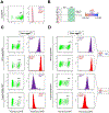
(A): Vero E6 cells transfected with EYFP and stained with either monoclonal (mAb) or polyclonal (pAb) antibodies against Trop-2. (B): Summary of the gating and signal acquisition strategy. (C,D): Left panels: dot plots of the experiments showing the YFP+ cells that were gated out for the subsequent analysis. Right panels: histogram plots showing the expression levels of membranous Trop-2 as measured by staining with an anti Trop-2 pAb (purple) or mAb (red). Ratios between mAb and pAb Mean Fluorescence Intensities are shown for each experimental group. Left, analysis on UGGT1+/+ cells.Right, analysis on UGGT1−/− cells.
Figure 7.
Figure 7.. Confocal microscopy of WT Trop-2 and Trop-2 Q118E, E227K and L186P mutants in Vero E6 cells.
Cellular localisation was assessed in UGGT1+/+ (left) and UGGT1−/− (right) Vero E6 cells by confocal microscopy. Cells were imaged upon co-transfection of WT Trop-2-EYFP (to track baseline localisation of the WT molecule) and WT/mutant Trop-2-mCherry fusion proteins (to track mutants with WT Trop-2). Mutant Trop-2 molecules were retained in intracellular compartments of the UGGT1+/+ cells (left), but reached the plasma membrane in UGGT1−/− cells. WT Trop-2-EYFP and WT Trop-2-mCherry co-localise at the plasma membrane independently on the presence or absence of UGGT1, as expected. White arrowheads: areas of membrane co-localisation. Red arrowheads: absence of co-localisation between WT Trop-2-EYFP and the mCherry Trop-2 mutant fusion proteins.

Update of

References

    1. Adams BM, Oster ME, and Hebert DN (2019). Protein Quality Control in the Endoplasmic Reticulum. Protein J. 38, 317–329. - PMC - PubMed
    1. Klausner RD, and Sitia R (1990). Protein degradation in the endoplasmic reticulum. Cell 62, 611–614. - PubMed
    1. Wiseman RL, Mesgarzadeh JS, and Hendershot LM (2022). Reshaping endoplasmic reticulum quality control through the unfolded protein response. Mol. Cell 82, 1477–1491. - PMC - PubMed
    1. Parenti G, Andria G, and Valenzano KJ (2015). Pharmacological Chaperone Therapy: Preclinical Development, Clinical Translation, and Prospects for the Treatment of Lysosomal Storage Disorders. Mol. Ther 23, 1138–1148. - PMC - PubMed
    1. Marinko JT, Huang H, Penn WD, Capra JA, Schlebach JP, and Sanders CR (2019). Folding and Misfolding of Human Membrane Proteins in Health and Disease: From Single Molecules to Cellular Proteostasis. Chem. Rev 119, 5537–5606. - PMC - PubMed