. 2024 Jan 25;15(1):739.
doi: 10.1038/s41467-024-45094-5.

LNP-RNA-engineered adipose stem cells for accelerated diabetic wound healing

Affiliations

LNP-RNA-engineered adipose stem cells for accelerated diabetic wound healing

Yonger Xue et al. Nat Commun. .

Abstract

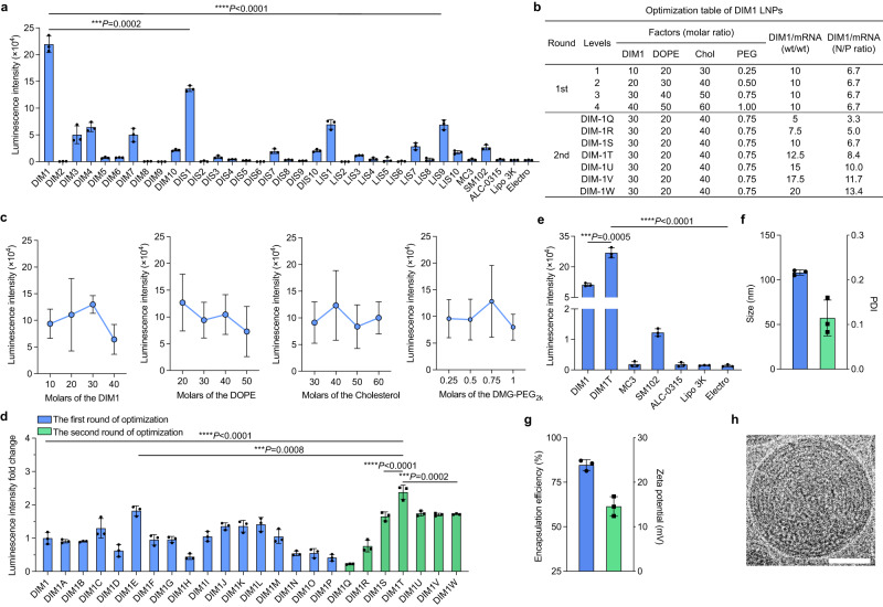
Adipose stem cells (ASCs) have attracted considerable attention as potential therapeutic agents due to their ability to promote tissue regeneration. However, their limited tissue repair capability has posed a challenge in achieving optimal therapeutic outcomes. Herein, we conceive a series of lipid nanoparticles to reprogram ASCs with durable protein secretion capacity for enhanced tissue engineering and regeneration. In vitro studies identify that the isomannide-derived lipid nanoparticles (DIM1T LNP) efficiently deliver RNAs to ASCs. Co-delivery of self-amplifying RNA (saRNA) and E3 mRNA complex (the combination of saRNA and E3 mRNA is named SEC) using DIM1T LNP modulates host immune responses against saRNAs and facilitates the durable production of proteins of interest in ASCs. The DIM1T LNP-SEC engineered ASCs (DS-ASCs) prolong expression of hepatocyte growth factor (HGF) and C-X-C motif chemokine ligand 12 (CXCL12), which show superior wound healing efficacy over their wild-type and DIM1T LNP-mRNA counterparts in the diabetic cutaneous wound model. Overall, this work suggests LNPs as an effective platform to engineer ASCs with enhanced protein generation ability, expediting the development of ASCs-based cell therapies.


Conflict of interest statement

Y.X., Y.Zhang., D.J.I., R.W., and Y.D. are inventors on a patent application (63/433,109) filed by The Ohio State University and Massachusetts Institute of Technology. The patent covers engineered ASCs and their uses in this work. Y.D. is a scientific advisory board member and holds equity in Arbor Biotechnologies and Sirnagen Therapeutics. Y.D. is a co-founder and holds equity in Immunanoengineering Therapeutics. D.J.I. and R.W. are scientific advisory board members and hold equity in Strand Therapeutics. Other authors declare no conflict of interest.

Figures

Fig. 1
Fig. 1. LNP-RNA-engineered adipose stem cell therapy to treat acute diabetic wounds.
a Illustration of LNP-engineered ASCs with enhanced protein-generating ability for diabetic wound healing. This illustration was created with BioRender.com and permitted for publication. b A representative synthetic route for sugar alcohol-derived ionizable lipids and structures of isomannide-derived ionizable lipids (DIM lipids).
Fig. 2
Fig. 2. Screening, optimization, and characterization of sugar alcohol-derived lipid nanoparticles.
a FLuc-mRNA delivery efficiency in primary murine ASCs represented as luminescence intensity. Data are from n = 3 biologically independent samples. b Table for the two rounds of DIM1/FLuc-mRNA LNP optimization. Chol cholesterol, PEG DMG-PEG2k. c Orthogonal assay to determine impact trends of each lipid component in DIM1 formulation at four levels. d Luminescence intensity fold changes of the two rounds of optimization. e Luminescence intensity of DIM1T LNPs and other control groups. f Hydrodynamic diameter (blue) and PDI (green) of DIM1T LNPs. g Encapsulation efficiency (blue) and zeta potential (green) of DIM1T LNPs. h Cryo-TEM image of DIM1T LNPs (scale bar = 50 nm). Data in h are representative images from n = 3 independent experiments. Data in a and cg are from n = 3 biologically independent samples and are presented as mean ± standard deviation (s.d.). One-way ANOVA followed by Dunnett’s multiple comparison test is used to determine the statistical significance and P values. ***P < 0.001, ****P < 0.0001.
Fig. 3
Fig. 3. Durable protein expression in ASCs.
a Kinetics of luminescence intensity DIM1T firefly-luciferase saRNA or mRNA LNPs treated ASCs after 2 days. b Luminescence intensity of DIM1T LNPs encapsulating FLuc-saRNA and E3 mRNA complex (SEC) at various saRNA/E3 mass ratios in ASCs after 48 h post-treatment. The total RNA dose is 50 ng/10000 cells. c Size distribution of DIM1T LNPs encapsulating FLuc mRNA, FLuc saRNA or FLuc SEC. d Kinetics of different DIM1T-RNAs LNPs in ASCs for 9 days. The statistical significance is analyzed using the two-tailed Student’s T test. e Surface expression levels of CD106, CD44, CD29, SCA-1, CD11b and CD45 on DS-ASCs. WT ASCs and isotype staining serve as controls. CD106 vascular cell adhesion molecule 1(VCAM-1), CD29 Integrin beta 1 (ITGB1), Sca-1 Stem Cell Antigen-1, CD11 Integrin alpha M (ITGAM). f Quantification of expression of each surface marker from e. g Cell viability of SEC-DIM1T LNPs in ASCs at various total RNA doses. h Uptake efficiency of DIM1T LNPs loaded with Alexa-Fluor 647 labeled RNAs in ASCs treated by various endocytic inhibitors. i CLSM images of ASCs stained with calcein alone or with DIM1T-LNPs (scale bar = 50 μm). Data in i are representative images from n = 3 independent experiments. Data in a, b, and dh are from n = 3 biologically independent samples and are presented as mean ± s.d. One-way ANOVA followed by Dunnett’s multiple comparison test is used to analyze the statistical significance. n.s. not significant, P > 0.05, *P < 0.05, **P < 0.01, ***P < 0.001, ****P < 0.0001.
Fig. 4
Fig. 4. DIM1T-HGF SEC LNPs-engineered ASCs accelerated wound healing in diabetic mice.
a Expression kinetics of HGF SEC delivered by DIM1T LNPs in ASCs. The conditioned medium was collected on Days 1, 2, 3, 5, 7, and 9. The protein level in the medium was analyzed using an ELISA kit. The statistical significance is analyzed using the two-tailed Student’s T-test. Data in a are from n = 3 biologically independent samples. b Representative digital image of the skin wounds of each group. c Relative wound size of vehicle controls, WT ASCs, HGF DM-ASCs, and HGF DS-ASCs. n = 10 for all groups. d Mean AUC of individual wounds of each group. n = 10 for all groups. e The complete wound closure time in vehicle controls, WT ASCs, HGF DM-ASCs, and HGF DS-ASCs. The significant differences in time to closure between groups are analyzed using the Log-rank test. **P < 0.01, ****P < 0.0001. f Representative Masson’s trichrome staining (MTS) of wounds on D18 for each group. h, j Representative CD31+ and αSMA+ immunofluorescence (IF) images of wounds on D18 for each group. g Quantification of the epidermal thickness of the wounds from f. i Quantification of CD31+ vessels per unit area on D18 from h. k Quantification of αSMA+ cells per unit area on D18 from j. Data in g, i, and k are from n = 10 biologically independent samples. All data are presented as mean ± s.d. Statistical significance and P values are analyzed by one-way ANOVA followed by Dunnett’s multiple comparison test. *P < 0.05, **P < 0.01, ***P < 0.001, ****P < 0.0001. Scale bars, 7 mm (b); 1 mm (f); 50 μm (h and j).
Fig. 5
Fig. 5. CXCL12-generating ASCs outperformed HGF counterparts in healing acute diabetic wounds.
a Expression kinetics of CXCL12 SEC delivered by DIM1T LNPs in ASCs. The statistical significance is analyzed using the two-tailed Student’s T-test. Data in a are from n = 3 biologically independent samples. b Representative digital image of the wounds of each group. c Relative wound size of vehicle controls, WT ASCs, HGF DS-ASCs, and CXCL12 DS-ASCs. n = 10 for all groups. d Mean AUC of individual wounds of each group. n = 10 wounds per group. e The complete wound closure time in vehicle controls, WT ASCs, HGF DS-ASCs, and CXCL12 DS-ASCs. The significant differences in time to closure between groups are analyzed using the Log-rank test. **P < 0.01. ****P < 0.0001. f Representative MTS and H&E images of wounds on D15 for each group. g Quantification of the epidermal thickness of the wounds on D15. h, j, l, n Representative CD31+, αSMA+, IL-6 and IL-10 IF images of wounds on D15 for each group. i, k, m, o Quantification of the CD31+ cells, the αSMA+ cells, the IL-6, and the IL-10. Data in g, i, k, m, and o are from n = 10 biologically independent samples. All data are presented as mean ± s.d. Statistical significance and P values are analyzed by one-way ANOVA followed by Dunnett’s multiple comparison test. *P < 0.05, **P < 0.01, ***P < 0.001, ****P < 0.0001. Scale bars, 7 mm (b); 1 mm (f); 50 μm (h, j, l, and n).

References

    1. Ankrum JA, Ong JF, Karp JM. Mesenchymal stem cells: immune evasive, not immune privileged. Nat. Biotechnol. 2014;32:252–260. doi: 10.1038/nbt.2816. - DOI - PMC - PubMed
    1. Wu X, et al. Mesenchymal stromal cell therapies: immunomodulatory properties and clinical progress. Stem Cell Res. Ther. 2020;11:345. doi: 10.1186/s13287-020-01855-9. - DOI - PMC - PubMed
    1. Xue Y, Baig R, Dong Y. Recent advances of biomaterials in stem cell therapies. Nanotechnology. 2022;33:132501. doi: 10.1088/1361-6528/ac4520. - DOI - PMC - PubMed
    1. Zhu Y, et al. Adipose‐derived stem cell: a better stem cell than BMSC. Cell Biochem. Funct. 2008;26:664–675. doi: 10.1002/cbf.1488. - DOI - PubMed
    1. Zuk PA, et al. Human adipose tissue is a source of multipotent stem cells. Mol. Biol. Cell. 2002;13:4279–4295. doi: 10.1091/mbc.e02-02-0105. - DOI - PMC - PubMed