. 2024 Jan 25;14(1):2202.
doi: 10.1038/s41598-024-52633-z.

Endoscopic and clinical features of gastric emphysema

Affiliations

Endoscopic and clinical features of gastric emphysema

Masaya Iwamuro et al. Sci Rep. .

Abstract

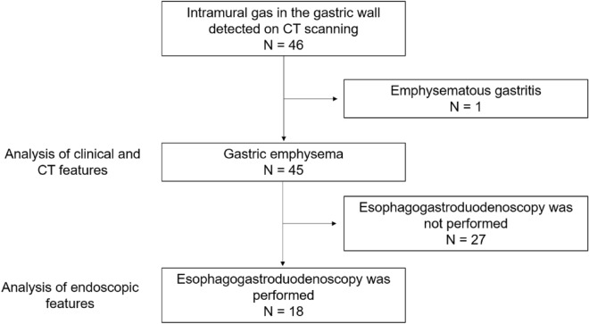
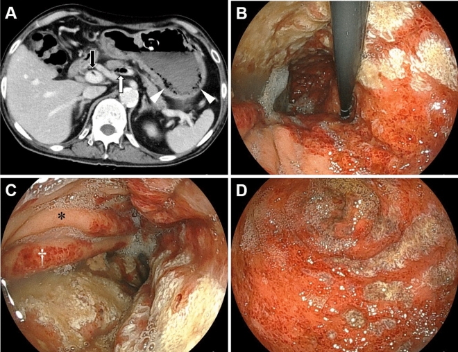
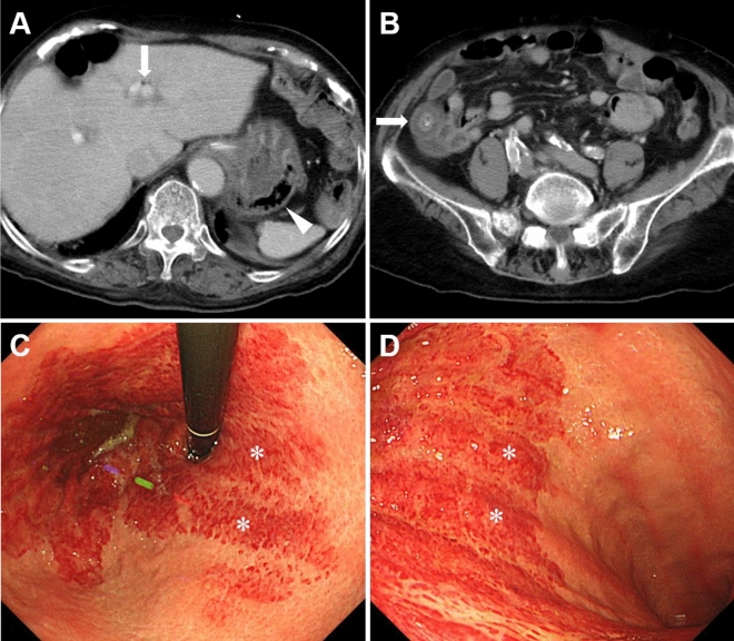
Gastric emphysema is characterized by the presence of intramural gas in the stomach without bacterial infection. Due to its rarity, most reports on gastric emphysema have been limited to single-case studies, and this condition's clinical and endoscopic features have not been thoroughly investigated. In this study, we analyzed 45 patients with gastric emphysema from 10 institutions and examined their characteristics, endoscopic features, and outcomes. The mean age at diagnosis of gastric emphysema in our study population (35 males and 10 females) was 68.6 years (range, 14-95 years). The top five underlying conditions associated with gastric emphysema were the placement of a nasogastric tube (26.7%), diabetes mellitus (20.0%), post-percutaneous endoscopic gastrostomy (17.8%), malignant neoplasms (17.8%), and renal failure (15.6%). Among the 45 patients, 42 were managed conservatively with fasting and administration of proton pump inhibitors. Unfortunately, seven patients died within 30 days of diagnosis, and 35 patients experienced favorable recoveries. The resolution of gastric emphysema was confirmed in 30 patients through computed tomography (CT) scans, with a mean duration of 17.1 ± 34.9 days (mean ± standard deviation [SD], range: 1-180 days) from the time of diagnosis to the disappearance of the gastric intramural gas. There were no instances of recurrence. Endoscopic evaluation was possible in 18 patients and revealed that gastric emphysema presented with features such as redness, erosion, coarse mucosa, and ulcers, with fewer mucosal injuries on the anterior wall (72.2%), a clear demarcation between areas of mucosal injury and intact mucosa (61.1%), and predominantly longitudinal mucosal injuries on the stomach folds (50.0%). This study is the first English-language report to analyze endoscopic findings in patients with gastric emphysema.


Conflict of interest statement

The authors declare no competing interests.

Figures

Figure 1
Figure 1
Study flow chart. CT computed tomography.
Figure 2
Figure 2
CT and endoscopic images of a 66-year-old male, currently undergoing chemotherapy for esophageal cancer, with a gastrostomy tube placement. The patient was asymptomatic, and during an endoscopic evaluation before esophageal cancer surgery, gastric mucosal injury was noted. On the same day, CT scans (A) revealed the presence of gas (white arrow) and thrombosis (black arrow) in the splenic to portal veins. Additionally, a gas-filled appearance was observed within the gastric wall (arrowheads). Esophagogastroduodenoscopy (BD; B, the gastric fornix and upper body; C, gastric body; D, gastric antrum) showed diffuse reddish erosions with white exudates extending from the fornix to the antrum. The boundary between the injured and non-injured mucosa was partially distinct (B). The anterior wall of the gastric body was intact (C, asterisk). Mucosal injury was predominant on the folds (C, dagger). CT computed tomography.
Figure 3
Figure 3
CT and endoscopic images of an 89-year-old female. The patient had a nasogastric tube inserted and a history of cerebral infarctions with internal carotid artery occlusion, chronic atrial fibrillation, aortic valve stenosis, and hypertrophic cardiomyopathy. She presented with a fever, and CT scans revealed portal venous gas (A, arrow), intramural gas in the stomach (A, arrowhead), and wall thickening of the ascending colon (B, arrow). Esophagogastroduodenoscopy (C,D) showed a circumferential erythematous erosion in the gastric fornix to the upper body. The boundary between the erythematous area and the intact mucosa was clear. The erythematous erosions were predominant on the convex part of the gastric folds and tended to be longitudinal (C,D, asterisks). CT computed tomography.

References

    1. López-Medina G, Castillo Díaz de León R, Heredia-Salazar AC, Hernández-Salcedo DR. Gastric emphysema a spectrum of pneumatosis intestinalis: A case report and literature review. Case Rep. Gastrointest. Med. 2014;2014:891360. - PMC - PubMed
    1. Soon MS, Yen HH, Soon A, Lin OS. Endoscopic ultrasonographic appearance of gastric emphysema. World J. Gastroenterol. 2005;11:1719–1721. doi: 10.3748/wjg.v11.i11.1719. - DOI - PMC - PubMed
    1. Shipman PJ, Drury P. Emphysematous gastritis: Case report and literature review. Australas. Radiol. 2001;45:64–66. doi: 10.1046/j.1440-1673.2001.00878.x. - DOI - PubMed
    1. Al-Jundi W, Shebl A. Emphysematous gastritis: Case report and literature review. Int. J. Surg. 2008;6:e63–e66. doi: 10.1016/j.ijsu.2007.02.007. - DOI - PubMed
    1. Loi TH, See JY, Diddapur RK, Issac JR. Emphysematous gastritis: A case report and a review of literature. Ann. Acad. Med. Singap. 2007;36:72–73. doi: 10.47102/annals-acadmedsg.V36N1p72. - DOI - PubMed