

Comparison of Widefield OCT Angiography Features Between Severe Non-Proliferative and Proliferative Diabetic Retinopathy
- PMID: 38273048
- PMCID: PMC10853160
- DOI: 10.1007/s40123-024-00886-2
Comparison of Widefield OCT Angiography Features Between Severe Non-Proliferative and Proliferative Diabetic Retinopathy
Abstract
Introduction: There is a high and ever-increasing global prevalence of diabetic retinopathy (DR) and invasive imaging techniques are often required to confirm the presence of proliferative disease. The aim of this study was to explore the images of a rapid and non-invasive technique, widefield optical coherence tomography angiography (OCT-A), to study differences between patients with severe non-proliferative and proliferative DR (PDR).
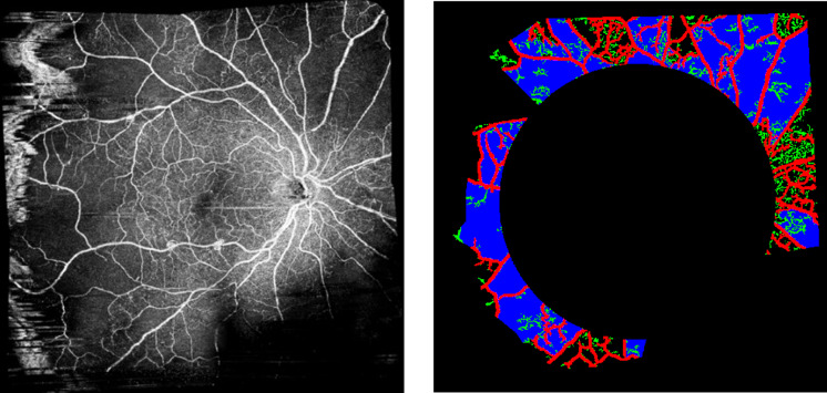
Methods: We conducted an observational longitudinal study from November 2022 to March 2023. We recruited 75 patients who were classified into a proliferative group (28 patients) and severe non-proliferative group (47 patients). Classification was done by specialist clinicians who had full access to any multimodal imaging they required to be confident of their diagnosis, including fluorescein angiography. For all patients, we performed single-shot 4 × 4 and 10 × 10 mm (widefield) OCT-A imaging and when possible, the multiple images required for mosaic 17.5 × 17.5 mm (ultra widefield) OCT-A imaging. We assessed the frequency with which proliferative disease was identifiable solely from these OCT-A images and used custom-built MATLAB software to analyze the images and determine computerized metrics such as density and intensity of vessels, foveal avascular zone, and ischemic areas.
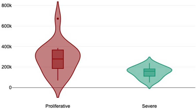
Results: On clinically assessing the OCT-A 10 × 10 fields, we were only able to detect new vessels in 25% of known proliferative images. Using ultra-widefield mosaic images, however, we were able to detect new vessels in 100% of PDR patients. The image analysis metrics of 4 × 4 and 10 × 10 mm images did not show any significant differences between the two clinical groups. For mosaics, however, there were significant differences in the capillary density in patients with PDR compared to severe non-PDR (9.1% ± 1.9 in the PDR group versus 11.0% ± 1.9 for severe group). We also found with mosaics a significant difference in the metrics of ischemic areas; average area of ischemic zones (253,930.1 ± 108,636 for the proliferative group versus 149,104.2 ± 55,101.8 for the severe group.
Conclusions: Our study showed a high sensitivity for detecting PDR using only ultra-widefield mosaic OCT-A imaging, compared to multimodal including fluorescein angiography imaging. It also suggests that image analysis of aspects such as ischemia levels may be useful in identifying higher risk groups as a warning sign for future conversion to neovascularization.
Keywords: Fluorescein angiography; Image analysis; New vessels; OCT-A; OCT-A metrics; Proliferative diabetic retinopathy; Retinal ischemia; Severe diabetic retinopathy.
© 2024. The Author(s).
Conflict of interest statement
Ines Drira, Maha Noor, Amy Stone, Yvonne D’Souza, Binu John, and Orlaith McGrath do not have any financial disclosures relevant for this article. Tariq Aslam has received grants/speaker fees from Novartis, Bayer, Roche, Heidelberg, Topcon, and Canon. Praveen J. Patel is a consultant to Bayer UK and Roche UK.
Figures

References
-
- Diabetes. Available from: https://www.who.int/health-topics/diabetes#tab=tab_1
-
- National Diabetes Statistics Report. Available from: https://www.cdc.gov/diabetes/data/statistics-report/index.html
LinkOut - more resources
Full Text Sources
