. 2024 Jan 25;7(1):130.
doi: 10.1038/s42003-023-05757-3.

KLF15 transcriptionally activates LINC00689 to inhibit colorectal cancer development

Affiliations

KLF15 transcriptionally activates LINC00689 to inhibit colorectal cancer development

Yan Cao et al. Commun Biol. .

Abstract

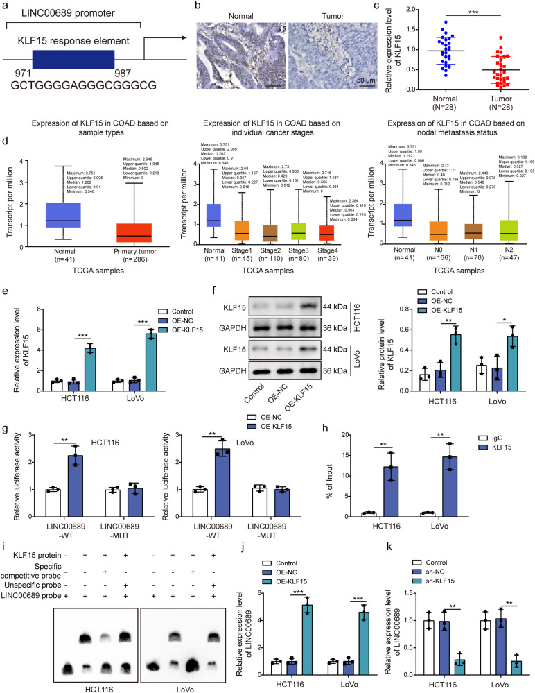
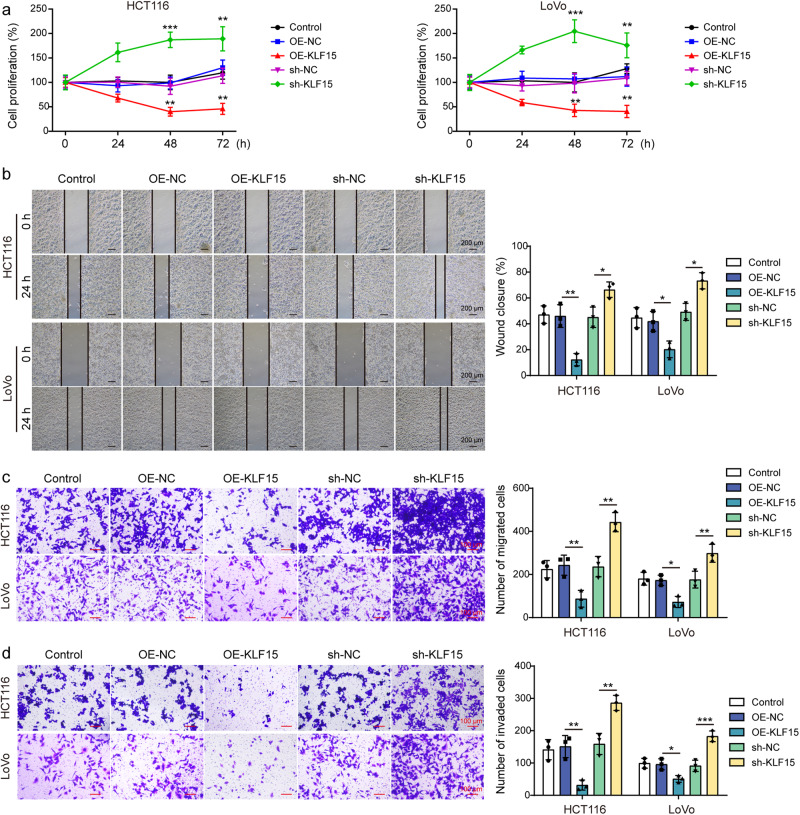
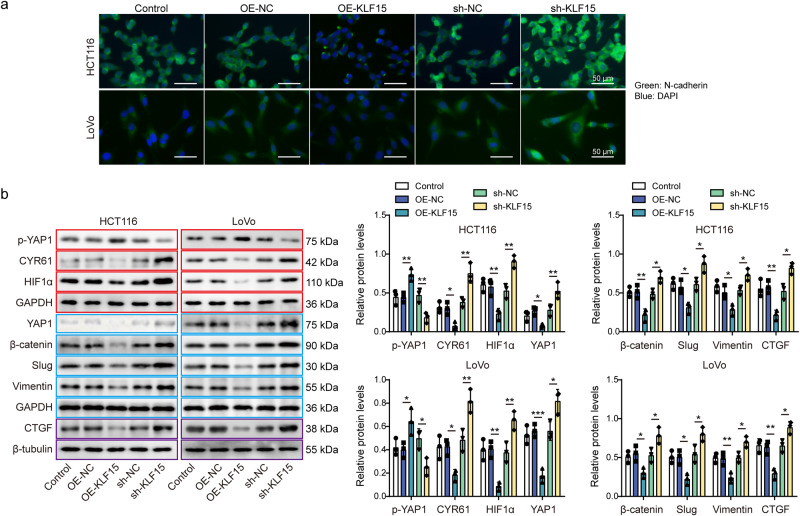
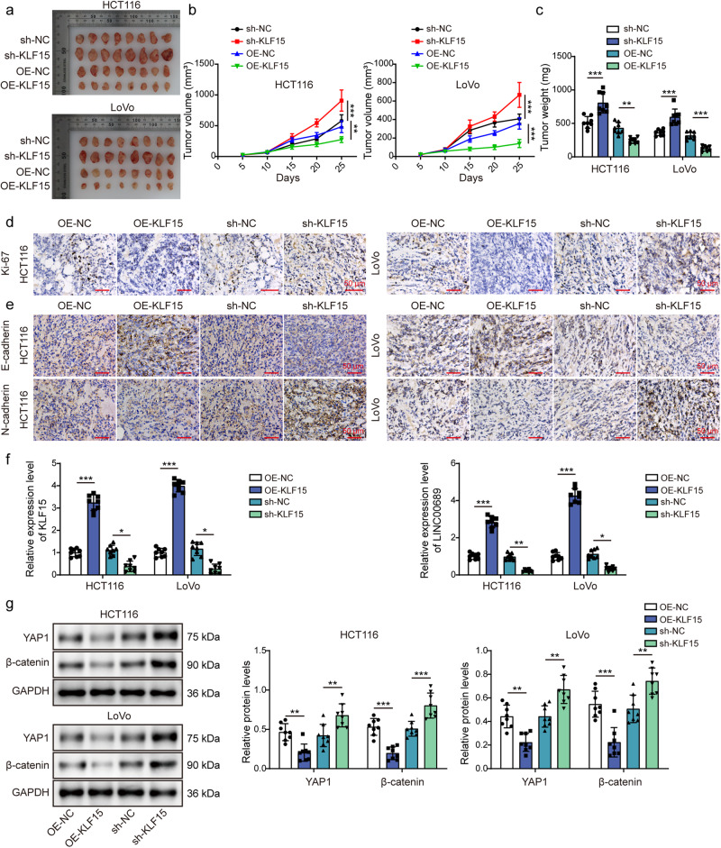
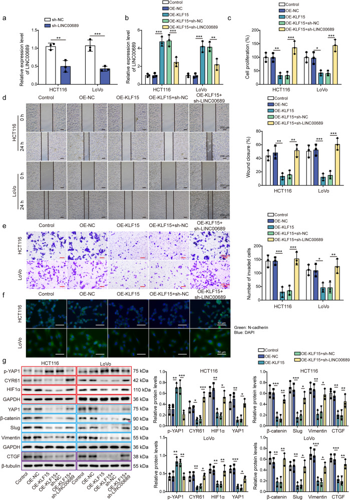
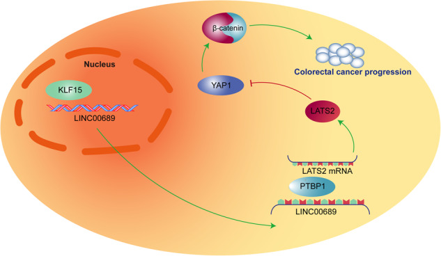
Colorectal cancer is a grievous health concern, we have proved long non-coding RNA LINC00689 is considered as a potential diagnosis biomarker for colorectal cancer, and it is necessary to further investigate its upstream and downstream mechanisms. Here, we show that KLF15, a transcription factor, exhibits the reduced expression in colorectal cancer. KLF15 suppresses the proliferative and metastatic capacities of colorectal cancer cells both in vitro and in vivo by transcriptionally activating LINC00689. Subsequently, LINC00689 recruits PTBP1 protein to enhance the stability of LATS2 mRNA in the cytoplasm. This stabilization causes the suppression of the YAP1/β-catenin pathway and its target downstream genes. Our findings highlight a regulatory network involving KLF15, LINC00689, PTBP1, LATS2, and the YAP1/β-catenin pathway in colorectal cancer, shedding light on potential therapeutic targets for colorectal cancer therapy.


Conflict of interest statement

The authors declare no competing interests.

Figures

Fig. 1
Fig. 1. KLF15 was downregulated in CRC and positively regulated LINC00689 level through binding to its promoter.
a hTFtarget database predicted the binding sites between transcription factor KLF15 and LINC00689 promoter. b, c Immunohistochemistry and RT-qPCR (n = 28) assays measured KLF15 levels in CRC tissues and adjacent non-tumor tissues. Scale bar: 50 μm. d UALCAN database indicated KLF15 relative levels in normal individuals and colon adenocarcinoma (COAD) samples of different clinicopathological features. The variables are sample types, individual cancer stages, and nodal metastasis status. The maximum, upper quartile, median, lower quartile, and minimum values were provided in (d). e, f The transfection efficiency of OE-KLF15 vector was evaluated by RT-qPCR and western blotting in HCT116 and LoVo cells. GAPDH was used as loading control for KLF15 in western blotting. g Luciferase reporter, (h) CHIP and (i) EMSA assays were used to analyze the interaction between KLF15 protein and LINC00689 promoter. j, k The relative expression of LINC00689 was measured by RT-qPCR after transfecting with OE-KLF15 or sh-KLF15 vector. n = 28 biologically independent samples in clinical results. n = 3 biologically independent experiments in in vitro results. Data were presented as mean ± SD except for d, *P < 0.05, **P < 0.01, and ***P < 0.001. KLF15 kruppel-like factor 15, OE overexpression, NC negative control, WT wild type, MUT mutant.
Fig. 2
Fig. 2. KLF15 inhibited the proliferative, migratory, and invasive abilities of CRC cells.
HCT116 and LoVo cells were transfected with OE-KLF15 or sh-KLF15 vector, (a) proliferation, (b, c) migration, and (d) invasion abilities of treated cells were assessed by CCK-8, wound healing, and transwell assays, respectively. Scale bar: 200 μm in (b) and 100 μm in (c/d). n = 3 biologically independent experiments in in vitro results. Data were presented as mean ± SD, *P < 0.05, **P < 0.01, and ***P < 0.001. KLF15 kruppel-like factor 15, OE overexpression, NC negative control.
Fig. 3
Fig. 3. KLF15 inhibited the expression of N-cadherin and YAP1/β-catenin signaling pathway in CRC cells.
a Immunofluorescence detection of N-cadherin expression in treated cells. Green, N-cadherin; Blue, DAPI. Scale bar: 50 μm. b p-YAP1 (S127), YAP1, β-catenin, slug, vimentin, CTGF, CYR61, and HIF1α levels in treated cells were measured by western blotting. β-tubulin was used as loading control for CTGF, and GAPDH was used as loading control for other proteins. Red, blue, or purple outlines denote bands that were derived from the same blot. n = 3 biologically independent experiments in in vitro results. Data were presented as mean ± SD, *P < 0.05, **P < 0.01, and ***P < 0.001. KLF15 kruppel-like factor 15, OE overexpression, NC negative control, YAP1 yes-associated protein 1, CTGF connective tissue growth factor, CYR61 cysteine-rich angiogenic inducer 61, HIF1α hypoxia inducible factor 1α.
Fig. 4
Fig. 4. Overexpression of KLF15 inhibited tumor growth in nude mice.
HCT116 and LoVo cells overexpressed or silenced KLF15 were injected into xenograft nude mice. After 25 days, mice were executed and tumor tissues were collected for subsequent experiments. a Images of tumors in different groups. b, c Quantification of tumor volume and weight in different groups. d, e The expression levels of Ki-67, E-cadherin, and N-cadherin in tumor tissues were measured by immunohistochemistry. Scale bar: 50 μm. f RT-qPCR evaluated KLF15 and LINC00689 levels in tumor tissues. g Western blotting determined YAP1 and β-catenin levels in tumor tissues. GAPDH was used as loading control in western blotting. n = 8 biologically independent animals. Data were presented mean ± SD, *P < 0.05, **P < 0.01, and ***P < 0.001. KLF15 kruppel-like factor 15, OE overexpression, NC negative control, YAP1 yes-associated protein 1.
Fig. 5
Fig. 5. LINC00689 knockout promoted the proliferative, migratory, and invasive abilities of CRC cells.
a Relative expression level of LINC00689 in wild type (WT) and LINC00689-KO CRC cells. The (b) proliferation, (c) migration, and (d) invasion abilities of WT and LINC00689-KO CRC cells were assessed by CCK-8, wound healing, and transwell assays, respectively. Scale bar: 200 μm in (c) and 100 μm in (d). e YAP1, β-catenin, slug, and vimentin levels in WT and LINC00689-KO CRC cells were measured by western blotting. GAPDH was used as loading control in western blotting. Red or blue outlines denote bands that were derived from the same blot. n = 3 biologically independent experiments in in vitro results. Data were presented as mean ± SD, *P < 0.05, **P < 0.01, and ***P < 0.001. YAP1 yes-associated protein 1, WT wild type, KO knockout.
Fig. 6
Fig. 6. Knockdown of LINC00689 abolished the function of KLF15 on CRC cell proliferation and metastasis, and activated YAP1/β-catenin pathway.
HCT116 and LoVo cells were co-transfected with sh-LINC00689 and OE-KLF15 vector. Then, a, b LINC00689 levels were detected by RT-qPCR. The (c) proliferation, (d) migration, and (e) invasion of treated cells were analyzed by CCK-8, wound healing, and transwell assays, respectively. Scale bar: 200 μm in (d) and 100 μm in (e). f The expression of N-cadherin was evaluated by immunofluorescence. Green, N-cadherin; Blue, DAPI. Scale bar: 50 μm. g Western blotting assessed p-YAP1 (S127), YAP1, β-catenin, slug, vimentin, CTGF, CYR61, and HIF1α levels. β-tubulin was used as loading control for CTGF, and GAPDH was used as loading control for other proteins. Red, blue, or purple outlines denote bands that were derived from the same blot. n = 3 biologically independent experiments in in vitro results. Data were presented as mean ± SD, *P < 0.05, **P < 0.01, and ***P < 0.001. KLF15 kruppel-like factor 15, OE overexpression, NC negative control, YAP1 yes-associated protein 1, CTGF connective tissue growth factor, CYR61 cysteine-rich angiogenic inducer 61, HIF1α hypoxia-inducible factor 1α.
Fig. 7
Fig. 7. LINC00689 recruited PTBP1 protein in CRC cells.
a RIP assay was conducted to confirm the binding of LINC00689 with five potential proteins (U2AF2, SRSF1, PTBP1, UPF1, and IGF2BP2) predicted by StarBase bioinformatic analysis. b The specific association of LINC00689 and PTBP1 protein was further evaluated using RNA pull-down assay. c The co-localization of LINC00689 and PTBP1 protein in the WT and LINC00689-KO HCT116 and LoVo cells was tested using FISH combined immunofluorescence staining. Scale bar: 10 μm. n = 3 biologically independent experiments in in vitro results. Data were presented as mean ± SD, *P < 0.05, **P < 0.01, and ***P < 0.001. U2AF2 U2 small nuclear RNA auxiliary factor 2, SRSF1 serine/arginine-rich splicing factor 1, PTBP1 polypyrimidine tract-binding protein 1, UPF1 upstream frameshift 1, IGF2BP2 insulin-like growth factor 2 mRNA binding protein 2.
Fig. 8
Fig. 8. LINC00689 recruited PTBP1 protein to regulate the stability of LATS2 mRNA in CRC cells.
a StarBase bioinformatic tool analyzed potential binding sites between PTBP1 and LATS2 mRNA. b The interaction between PTBP1 and LATS2 mRNA in HCT116 and LoVo cells was verified by RIP assay. c, d The protein (n = 14) and mRNA (n = 28) levels of LATS2 in CRC tissues and adjacent normal tissues were determined by western blotting and RT-qPCR. GAPDH was used as loading control in western blotting. e The relative expression of PTBP1 was detected in HCT116 and LoVo cells transfected with OE-PTBP1, sh-PTBP1#1, or sh-PTBP1#2 plasmid by RT-qPCR. f, g The mRNA and protein expression levels of LATS2 were measured by RT-qPCR and western blotting after overexpressing or knocking down PTBP1. GAPDH was used as loading control in western blotting. h LATS2 mRNA levels in HCT116 and LoVo cells co-transfected with OE-PTBP1+sh-LINC00689 were examined by RT-qPCR after treatment with 2 μg/mL act-D for 0, 4, 8, 12, and 24 h. n = 28 biologically independent samples in clinical results. n = 3 biologically independent experiments in in vitro results. Data were presented as mean ± SD, *P < 0.05, **P < 0.01, and ***P < 0.001. PTBP1 polypyrimidine tract-binding protein 1, LATS2 large tumor suppressor kinase 2, OE overexpression, NC negative control.
Fig. 9
Fig. 9. LINC00689 inhibited proliferation, migration, and invasion of CRC cells through upregulating LATS2.
a LINC00689, and (b) p-YAP1 (S127), YAP1, β-catenin, slug, vimentin, CTGF, CYR61, and HIF1α protein expression in CRC cells co-transfected with OE-LINC00689 and sh-LATS2 were examined by RT-qPCR and western blotting, respectively. β-tubulin was used as loading control for CTGF, and GAPDH was used as loading control for other proteins in western blotting. Red, blue, or purple outlines denote bands that were derived from the same blot. c CRC cell proliferative, (d) migratory, and (e) invasive abilities were evaluated after overexpressing LINC00689 and silencing LATS2 via CCK-8, wound healing, and transwell assays, respectively. Scale bar: 200 μm in d and 100 μm in e. f Immunofluorescence detection of N-cadherin expression after simultaneous overexpression of LINC00689 and knockdown of LATS2. Green, N-cadherin; Blue, DAPI. Scale bar: 50 μm. n = 3 biologically independent experiments in in vitro results. Data were presented as mean ± SD, *P < 0.05, **P < 0.01, and ***P < 0.001. LATS2 large tumor suppressor kinase 2, OE overexpression, NC negative control, YAP1 yes-associated protein 1, CTGF connective tissue growth factor, CYR61 cysteine-rich angiogenic inducer 61, HIF1α hypoxia inducible factor 1α.
Fig. 10
Fig. 10. Graphical abstract.
Transcription factor KLF15 was found to upregulate LINC00689 via binding to its promoter, then the upregulated LINC00689 could enhance the stability of LATS2 mRNA through interacting with PTBP1 protein, thereby inhibiting CRC development by repressing the activation of YAP1/β-catenin pathway.

References

    1. Weitz J, et al. Colorectal cancer. Lancet. 2005;365:153–165. doi: 10.1016/S0140-6736(05)17706-X. - DOI - PubMed
    1. Zhang C, et al. Histological tumor response to neoadjuvant chemotherapy correlates to immunoscore in colorectal cancer liver metastases patients. J Surg Oncol. 2021;124:1431–1441. doi: 10.1002/jso.26651. - DOI - PubMed
    1. Dekker E, Tanis PJ, Vleugels JLA, Kasi PM, Wallace MB. Colorectal cancer. Lancet. 2019;394:1467–1480. doi: 10.1016/S0140-6736(19)32319-0. - DOI - PubMed
    1. Wang KC, Chang HY. Molecular mechanisms of long noncoding RNAs. Mol. Cell. 2011;43:904–914. doi: 10.1016/j.molcel.2011.08.018. - DOI - PMC - PubMed
    1. Hou P, et al. Correction to: LINC00460/DHX9/IGF2BP2 complex promotes colorectal cancer proliferation and metastasis by mediating HMGA1 mRNA stability depending on m6A modification. J. Exp. Clin. Cancer Res. 2021;40:365. doi: 10.1186/s13046-021-02169-1. - DOI - PMC - PubMed

Publication types

MeSH terms

Substances