Review
. 2024 Jan 25;22(1):15.
doi: 10.1186/s12915-024-01824-1.

Mitochondrial genomes revisited: why do different lineages retain different genes?

Affiliations
Review

Mitochondrial genomes revisited: why do different lineages retain different genes?

Anzhelika Butenko et al. BMC Biol. .

Abstract

The mitochondria contain their own genome derived from an alphaproteobacterial endosymbiont. From thousands of protein-coding genes originally encoded by their ancestor, only between 1 and about 70 are encoded on extant mitochondrial genomes (mitogenomes). Thanks to a dramatically increasing number of sequenced and annotated mitogenomes a coherent picture of why some genes were lost, or relocated to the nucleus, is emerging. In this review, we describe the characteristics of mitochondria-to-nucleus gene transfer and the resulting varied content of mitogenomes across eukaryotes. We introduce a 'burst-upon-drift' model to best explain nuclear-mitochondrial population genetics with flares of transfer due to genetic drift.

Keywords: CoRR hypothesis; Endosymbiont gene transfer; Evolutionary cell biology; Mitochondrial DNA; Mitochondrial evolution; Mitochondrial mutation rates.


Conflict of interest statement

The authors declare that they have no competing interests.

Figures

Fig. 1
Fig. 1
Most lineages retain relatively stable mitochondrial genomic coding capacities. A cladogram starting with LECA depicts the differential evolution of mitochondrial genomic coding capacities in widely divergent eukaryotic lineages. Though exceptions to these trends are present in various groups, several lineages retain mitochondrial genome coding capacities typical for their clade. CII indicates retention of (some) complex II subunits; ccm indicates retention of subunits of the multicomponent bacteria-derived c-type cytochrome biogenesis system. Purple lineage: largest set of mitochondrial genes; orange lineages: retention of an intermediate number of mitochondrial genes; red lineages: retention of the ‘core set’ of mitochondrial genes only; black lineages: more extensive mitochondrial gene transfer and loss including transfer or loss of all ribosomal genes—usually contains fragmented rRNAs. Asterisks indicate lineages displaying large variations in mitochondrial gene content. For further information, see the main text
Fig. 2
Fig. 2
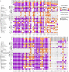
Mitochondria-to-nucleus gene transfer is relatively rare. Coulson plots showing the distribution of genes encoding components of small (A) and large (B) subunits of mitoribosomes, as well as electron transport chain (C) and other (D) proteins across mitochondrial and nuclear genomes of the representatives of major eukaryotic supergroups. Genes retained in the mitogenomes are depicted in purple, nucleus-encoded genes are in orange, and those lost or not detected are in white. Within each eukaryotic group, the species with identical gene distribution patterns were unified into one sector of a circle. Species with available genome and transcriptome assemblies are marked with back circles and triangles, respectively. The cladogram reflecting the phylogenetic relationships among major eukaryotic lineages is based on [9] and [10]. The bar charts at the top of the Coulson plots indicate the percentage of investigated taxa where the respective gene is encoded in mitochondrial (purple) and nuclear genome (orange) or lost/not detected. The numerical values above the bar charts correspond to the number of presumably independent mitochondrion-to-nucleus gene transfer events in the evolution of eukaryotes. Proteins predicted to possess a mitochondrial presequence by at least two out of three bioinformatic tools (MitoFates, TargetP, and TPpred3) are marked with cyan circles. The presence of a gene encoding cytochrome c heme-lyase in the nuclear genome is indicated with an asterisk over the ccmA gene charts. Coulson plots were produced with a Coulson plot generator [11]. For compact representation, some species were assigned numbers as follows: 1, Naegleria gruberi; 2, Naegleria fowleri; 3, Andalucia godoyi; 4, Reclinomonas americana; 5, Euglena gracilis; 6, Euglenozoa ‘SAG EU17/18’; 7, Diplonema papillatum; 8, Trypanosoma brucei; 9, Tsukubamonas globosa; 10, Heterolobosea sp. ‘BB2’; 11, Acrasis kona; 12, Pharyngomonas kirbyi; 13, Plasmodium falciparum; 14, Babesia microti; 15, Cyclospora cayetanensis; 16, Theileria annulata; 17, Toxoplasma gondii; 18, Phaeodactylum tricornutum; and 19, Thalassiosira pseudonana. The species abbreviations: Acar, Amphidinium carterae; Acas, Acanthamoeba castellanii; Ainv, Aphanomyces invadans; Amac, Allomyces macrogynus; Apac, Alexandrium pacificum; Aper, Acavomonas peruviana; Atwi, Ancoracysta twista; Blasto, Blastocystis sp.; Bmot, Brevimastigomonas motovehiculus; Bnat, Bigelowiella natans; Cbur, Cafeteria burkhardae; Cmar, Chattonella marina; Cmer, Cyanidioschyzon merolae; Cowc, Capsaspora owczarzaki; Cpar, Cyanophora paradoxa; Crei, Chlamydomonas reinhardtii; Ctob, Chrysochromulina tobinii; Cvel, Chromera velia; Cvie, Colponema vietnamica; Dbru, Dekkera bruxellensis; Ddis, Dictyostelium discoideum; Drot, Diphylleia rotans; Ehux, Emiliania huxleyi; Eten, Eimeria tenella; Falb, Fonticula alba; Ginc, Glaucocystis incrassata; Gsul, Galdieria sulphuraria; Gthe, Guillardia theta; Hand, Hemiselmis andersenii; Hema, Hematodinium sp.; Hmar, Hemiarma marina; Hsap, Homo sapiens; Lcau, Leucocytozoon caulleryi; Lmar, Leucocryptos marina; Maro, Marophrys sp.; MAST, marine stramenopile; Mbre, Monosiga brevicollis; Mcal, Malawimonas californiana; Mjak, Malawimonas jakobiformis; Mpol, Marchantia polymorpha; Mvir, Mesostigma viride; Noli, Nephroselmis olivacea; Nova, Nyctotherus ovalis; Nqua, Nibbleromonas quarantinus; Nsim, Nuclearia simplex; Ntab, Nicotiana tabacum; Omar, Oxyrrhis marina; Otri, Oxytricha trifallax; Pbil, Palpitomonas bilix; Pbra, Plasmodiophora brassicae; Perkma, Perkinsus marinus; Pico, Picozoa sp.; Pmar, Paracercomonas marina; Pmin, Pedinomonas minor; Ppro, Pycnococcus provasolii; Ppur, Porphyra purpurea; Pwic, Prototheca wickerhamii; Rsal, Rhodomonas salina; Scer, Saccharomyces cerevisiae; Stro, Strombidium sp.; Taur, Thraustochytrium aureum; TelT, Telonemid sp.; The, Tetrahymena thermophila; Ttra, Thecamonas trahens; Vbra, Vitrella brassicaformis; and Vver, Vermamoeba vermiformis
Fig. 3
Fig. 3
Obstacles to functional mitochondria-to-nucleus gene transfer. Subsequent steps in the transfer of mitochondrial (mt) genes to the nuclear genomes are indicated with (numbered) grey arrows. Obstacles to transfer are marked by letters (A–H) and arrows (CoRR hypothesis: red; constraints hypothesis: magenta). Genetic material can be transferred from the mitochondria to the nuclei as DNA or cDNA (1) during fission/fusion events, mitochondrial lysis or mitophagy, the transfer process being facilitated by organelle proximity and vacuole formation, protecting DNA fragments from cytoplasmic nucleases. Entrenched mitochondrial gene regulation can be a barrier to transfer. A specific case of regulation of expression by redox sensors and redox response regulators forms the crux of the CoRR hypothesis. Gene transfer in the opposite direction (nucleus-to-mitochondrial genome (2)) is extremely rare (so far, only demonstrated in corals and plants). Upon (c)DNA transfer, integration into a suitable genome locus (B) without disrupting essential genes or causing genome instability has to occur. Some genes will gain mitochondrial targeting signals (orange segments) from other nuclear genes (C) or formed de novo. The newly transferred gene should gain regulatory elements (green dots) enabling efficient expression (D) or be transcribed polycistronically with a nuclear gene. The process of codon optimization might contribute to establishing optimal expression levels of the now nucleus-encoded gene (D). For some organisms, mitochondrial RNA editing/deviations of the genetic code might represent extra obstacles to effective gene transfer (D). Upon successful completion of the steps mentioned, mRNA is synthesized and exported to the cytoplasm (3), where proteins are synthesized (4) on cytosolic ribosomes (olive green circles). Proteins with highly hydrophobic transmembrane domains, >  ~ 120 amino acids (length threshold for proteins to be recognized by the signal recognition particle), would thus be co-translationally miss-targeted to the ER (E). Newly synthesized proteins might be degraded by cytoplasmic peptidases (F) or bind chaperones (5) and be directed to mitochondria. Proteins enter mitochondria using a pre-sequence mediated pathway involving TOM and TIM23 complexes (6), with subsequent cleavage of pre-sequences by mitochondrial processing peptidase (7), or via other mechanisms (8). High protein hydrophobicity might represent a significant barrier to traversing the mitochondrial membranes (G). Following a successful transport into the mitochondria, proteins assume native conformations (9) and in some cases are incorporated into their respective protein complexes (10). Protein complex assembly processes normally involve highly ordered sets of steps, often requiring co-translational incorporation of subunits, potentially representing an additional barrier (H) for functional gene transfer to the nucleus
Fig. 4
Fig. 4
Burst-upon-drift (BUD) model: Small population sizes and high mitochondrial mutation rates can lead to fixation of slightly deleterious mitochondria-to-nucleus gene transfers. Path 1→ 2 represents mitochondria to nucleus transfer by adaptive mechanisms. Path 1 → 3 → 4 → 5 represents neutral transfers via the BUD model. (1) A mitogene is transferred to the nucleus (nu) and is transcribed, translated, and effectively targeted to the mitochondria (red cell in between orange cells). (2) The newly nuclear mitogene (nu) is beneficial and sweeps to fixation in a population due to natural selection, while the mitochondrial mitogene (mito) is lost because of bioenergetic benefit. New adaptations (nu*) will evolve in response to the new genomic location of the previous mitogene. (3) If the newly nuclear mitogene is neutral or mildly detrimental, the transfer can be fixed in the population by drift. In this situation, it is possible that the (mito)gene acquires moderate mutations leading to the sub-functionalization of the gene duplicates and their subsequent retention. (4) Loss of the mitogene may be fixed by drift if the mitochondrial mutation rate is high in a small population. In certain situations, this can occur even though there is a fitness cost caused by retaining only the nuclear mitogene. In these cases, several genes may transfer in quick succession leading to many fewer genes being encoded in the mitochondrial genome (black cells). (5) After the recovery from the population bottleneck, new adaptations (nu*) will evolve in response to the new genomic location of the previous mitogene. Ovals depict individual cells; colour changes of contours reflect changed cells (when compared with cells from a previous step). The colour code is consistent with the lineages in Fig. 1

Similar articles

Cited by

References

    1. Roger AJ, Munoz-Gomez SA, Kamikawa R. The origin and diversification of mitochondria. Curr Biol. 2017;27(21):R1177–R1192. doi: 10.1016/j.cub.2017.09.015. - DOI - PubMed
    1. Strassert JFH, Irisarri I, Williams TA, Burki F. A molecular timescale for eukaryote evolution with implications for the origin of red algal-derived plastids. Nat Commun. 2021;12(1):1879. doi: 10.1038/s41467-021-22044-z. - DOI - PMC - PubMed
    1. Al Jewari C, Baldauf SL. An excavate root for the eukaryote tree of life. Sci Adv. 2023;9(17):eade4973. doi: 10.1126/sciadv.ade4973. - DOI - PMC - PubMed
    1. Burger G, Gray MW, Forget L, Lang BF. Strikingly bacteria-like and gene-rich mitochondrial genomes throughout jakobid protists. Genome Biol Evol. 2013;5(2):418–438. doi: 10.1093/gbe/evt008. - DOI - PMC - PubMed
    1. Janouškovec J, Tikhonenkov DV, Burki F, Howe AT, Rohwer FL, Mylnikov AP, Keeling PJ. A new lineage of eukaryotes illuminates early mitochondrial genome reduction. Curr Biol. 2017;27(23):3717–3724.e3715. doi: 10.1016/j.cub.2017.10.051. - DOI - PubMed

LinkOut - more resources