

Development of a generative deep learning model to improve epiretinal membrane detection in fundus photography
- PMID: 38273286
- PMCID: PMC10811871
- DOI: 10.1186/s12911-024-02431-4
Development of a generative deep learning model to improve epiretinal membrane detection in fundus photography
Abstract
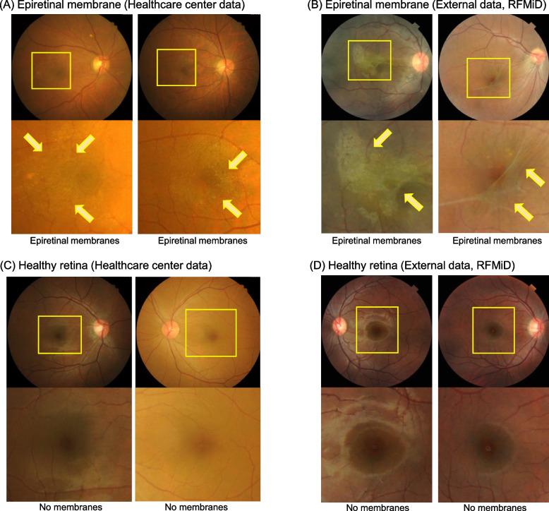
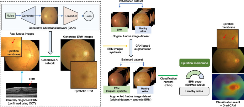
Background: The epiretinal membrane (ERM) is a common retinal disorder characterized by abnormal fibrocellular tissue at the vitreomacular interface. Most patients with ERM are asymptomatic at early stages. Therefore, screening for ERM will become increasingly important. Despite the high prevalence of ERM, few deep learning studies have investigated ERM detection in the color fundus photography (CFP) domain. In this study, we built a generative model to enhance ERM detection performance in the CFP.
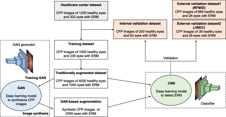
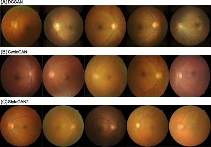
Methods: This deep learning study retrospectively collected 302 ERM and 1,250 healthy CFP data points from a healthcare center. The generative model using StyleGAN2 was trained using single-center data. EfficientNetB0 with StyleGAN2-based augmentation was validated using independent internal single-center data and external datasets. We randomly assigned healthcare center data to the development (80%) and internal validation (20%) datasets. Data from two publicly accessible sources were used as external validation datasets.
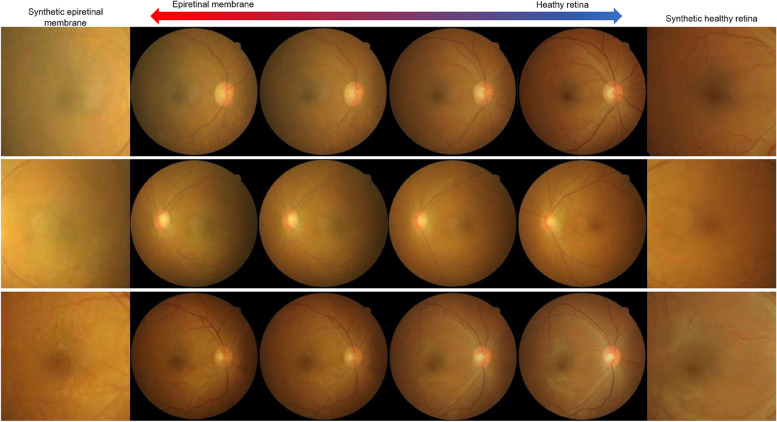
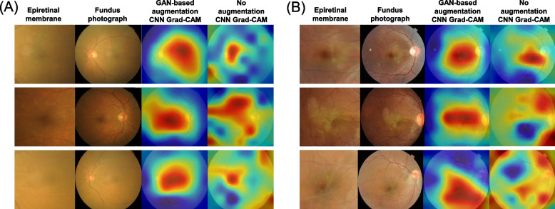
Results: StyleGAN2 facilitated realistic CFP synthesis with the characteristic cellophane reflex features of the ERM. The proposed method with StyleGAN2-based augmentation outperformed the typical transfer learning without a generative adversarial network. The proposed model achieved an area under the receiver operating characteristic (AUC) curve of 0.926 for internal validation. AUCs of 0.951 and 0.914 were obtained for the two external validation datasets. Compared with the deep learning model without augmentation, StyleGAN2-based augmentation improved the detection performance and contributed to the focus on the location of the ERM.
Conclusions: We proposed an ERM detection model by synthesizing realistic CFP images with the pathological features of ERM through generative deep learning. We believe that our deep learning framework will help achieve a more accurate detection of ERM in a limited data setting.
Keywords: Deep learning; Epiretinal membrane; Fundus photography; Generative adversarial net.
© 2024. The Author(s).
Conflict of interest statement
IHR and JKK are directors of VISUWORKS, and own company stock. IHR serves on the Advisory Board for Carl Zeiss Meditec AG and Avellino Lab USA/MAB for Avellino Lab Korea. TKY is an employee of VISUWORKS and received a salary or stock as part of the standard compensation package. The remaining authors declare no conflicts of interest.
Figures

Similar articles
-
Comparison of MultiColor fundus imaging and colour fundus photography in the evaluation of epiretinal membrane.Acta Ophthalmol. 2019 Jun;97(4):e533-e539. doi: 10.1111/aos.13978. Epub 2018 Nov 22. Acta Ophthalmol. 2019. PMID: 30565886
-
Multicolor Scanning Laser Ophthalmoscopy Strengthens Surgeons' Preoperative Decision-Making and Intraoperative Performance on Epiretinal Membrane.Transl Vis Sci Technol. 2020 Dec 18;9(13):36. doi: 10.1167/tvst.9.13.36. eCollection 2020 Dec. Transl Vis Sci Technol. 2020. PMID: 33384890 Free PMC article. Clinical Trial.
-
Epiretinal Membrane Detection at the Ophthalmologist Level using Deep Learning of Optical Coherence Tomography.Sci Rep. 2020 May 21;10(1):8424. doi: 10.1038/s41598-020-65405-2. Sci Rep. 2020. PMID: 32439844 Free PMC article.
-
Deep learning can generate traditional retinal fundus photographs using ultra-widefield images via generative adversarial networks.Comput Methods Programs Biomed. 2020 Dec;197:105761. doi: 10.1016/j.cmpb.2020.105761. Epub 2020 Sep 16. Comput Methods Programs Biomed. 2020. PMID: 32961385
-
Surgical Treatment of Epiretinal Membrane.Acta Med Okayama. 2021 Aug;75(4):403-413. doi: 10.18926/AMO/62378. Acta Med Okayama. 2021. PMID: 34511606 Review.
Cited by
-
The Role of Artificial Intelligence in Epiretinal Membrane Care: A Scoping Review.Ophthalmol Sci. 2024 Dec 20;5(4):100689. doi: 10.1016/j.xops.2024.100689. eCollection 2025 Jul-Aug. Ophthalmol Sci. 2024. PMID: 40182981 Free PMC article. Review.
-
Classification of Diabetic Retinopathy Disease Levels by Extracting Spectral Features Using Wavelet CNN.Diagnostics (Basel). 2024 May 24;14(11):1093. doi: 10.3390/diagnostics14111093. Diagnostics (Basel). 2024. PMID: 38893619 Free PMC article.
-
Development of oculomics artificial intelligence for cardiovascular risk factors: A case study in fundus oculomics for HbA1c assessment and clinically relevant considerations for clinicians.Asia Pac J Ophthalmol (Phila). 2024 Jul-Aug;13(4):100095. doi: 10.1016/j.apjo.2024.100095. Epub 2024 Aug 28. Asia Pac J Ophthalmol (Phila). 2024. PMID: 39209216 Free PMC article.
-
Discriminative, generative artificial intelligence, and foundation models in retina imaging.Taiwan J Ophthalmol. 2024 Nov 28;14(4):473-485. doi: 10.4103/tjo.TJO-D-24-00064. eCollection 2024 Oct-Dec. Taiwan J Ophthalmol. 2024. PMID: 39803410 Free PMC article. Review.
References
MeSH terms
LinkOut - more resources
Full Text Sources
Miscellaneous
