

Collagen in the central nervous system: contributions to neurodegeneration and promise as a therapeutic target
- PMID: 38273335
- PMCID: PMC10809576
- DOI: 10.1186/s13024-024-00704-0
Collagen in the central nervous system: contributions to neurodegeneration and promise as a therapeutic target
Abstract
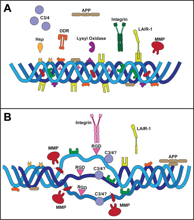
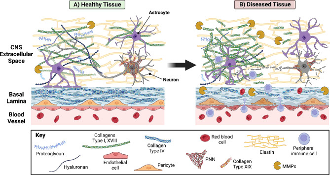
The extracellular matrix is a richly bioactive composition of substrates that provides biophysical stability, facilitates intercellular signaling, and both reflects and governs the physiological status of the local microenvironment. The matrix in the central nervous system (CNS) is far from simply an inert scaffold for mechanical support, instead conducting an active role in homeostasis and providing broad capacity for adaptation and remodeling in response to stress that otherwise would challenge equilibrium between neuronal, glial, and vascular elements. A major constituent is collagen, whose characteristic triple helical structure renders mechanical and biochemical stability to enable bidirectional crosstalk between matrix and resident cells. Multiple members of the collagen superfamily are critical to neuronal maturation and circuit formation, axon guidance, and synaptogenesis in the brain. In mature tissue, collagen interacts with other fibrous proteins and glycoproteins to sustain a three-dimensional medium through which complex networks of cells can communicate. While critical for matrix scaffolding, collagen in the CNS is also highly dynamic, with multiple binding sites for partnering matrix proteins, cell-surface receptors, and other ligands. These interactions are emerging as critical mediators of CNS disease and injury, particularly regarding changes in matrix stiffness, astrocyte recruitment and reactivity, and pro-inflammatory signaling in local microenvironments. Changes in the structure and/or deposition of collagen impact cellular signaling and tissue biomechanics in the brain, which in turn can alter cellular responses including antigenicity, angiogenesis, gliosis, and recruitment of immune-related cells. These factors, each involving matrix collagen, contribute to the limited capacity for regeneration of CNS tissue. Emerging therapeutics that attempt to rebuild the matrix using peptide fragments, including collagen-enriched scaffolds and mimetics, hold great potential to promote neural repair and regeneration. Recent evidence from our group and others indicates that repairing protease-degraded collagen helices with mimetic peptides helps restore CNS tissue and promote neuronal survival in a broad spectrum of degenerative conditions. Restoration likely involves bolstering matrix stiffness to reduce the potential for astrocyte reactivity and local inflammation as well as repairing inhibitory binding sites for immune-signaling ligands. Facilitating repair rather than endogenous replacement of collagen degraded by disease or injury may represent the next frontier in developing therapies based on protection, repair, and regeneration of neurons in the central nervous system.
Keywords: Alzheimer’s Disease; Collagen; Collagen mimetic peptide; Extracellular matrix; Glaucoma; Neuro-regeneration; Neuro-replacement; Neurodegeneration; Neurovascular coupling.
© 2024. The Author(s).
Conflict of interest statement
Not applicable.
Figures
References
-
- Hussey GS, Dziki JL, Badylak SF. Extracellular matrix-based materials for regenerative medicine. Nat Reviews Mater. 2018;3(7):159–73. doi: 10.1038/s41578-018-0023-x. - DOI
Publication types
MeSH terms
Substances
Grants and funding
LinkOut - more resources
Full Text Sources
