. 2024 Jan 23;18(1):014103.
doi: 10.1063/5.0181287. eCollection 2024 Jan.

Machine learning classification of cellular states based on the impedance features derived from microfluidic single-cell impedance flow cytometry

Affiliations

Machine learning classification of cellular states based on the impedance features derived from microfluidic single-cell impedance flow cytometry

Jian Wei et al. Biomicrofluidics. .

Abstract

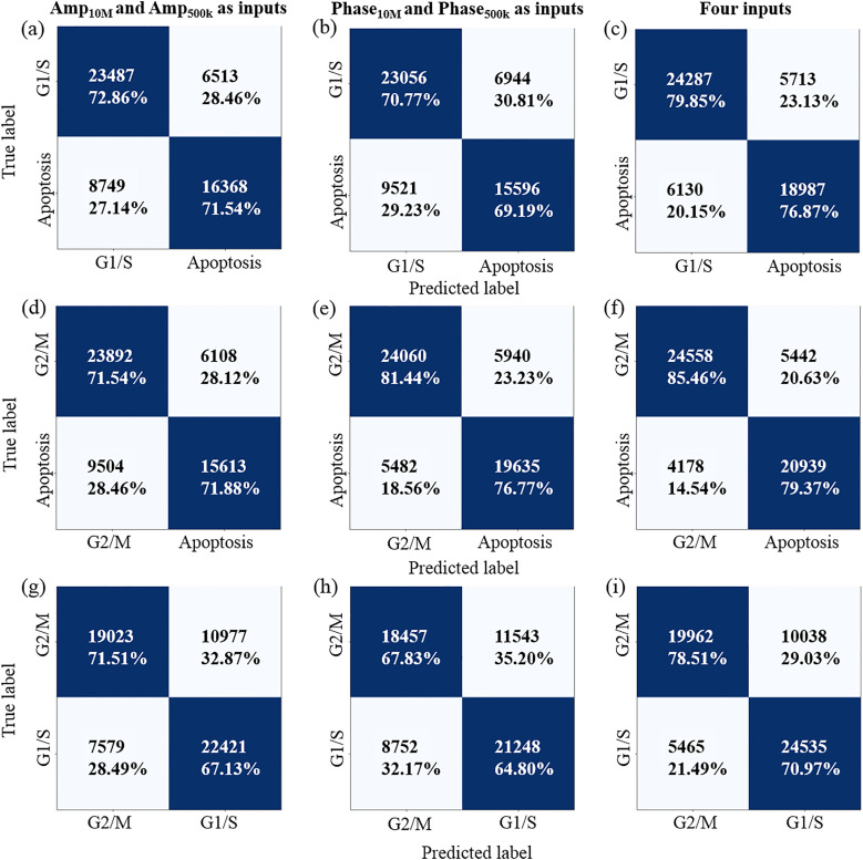
Mitosis is a crucial biological process where a parental cell undergoes precisely controlled functional phases and divides into two daughter cells. Some drugs can inhibit cell mitosis, for instance, the anti-cancer drugs interacting with the tumor cell proliferation and leading to mitosis arrest at a specific phase or cell death eventually. Combining machine learning with microfluidic impedance flow cytometry (IFC) offers a concise way for label-free and high-throughput classification of drug-treated cells at single-cell level. IFC-based single-cell analysis generates a large amount of data related to the cell electrophysiology parameters, and machine learning helps establish correlations between these data and specific cell states. This work demonstrates the application of machine learning for cell state classification, including the binary differentiations between the G1/S and apoptosis states and between the G2/M and apoptosis states, as well as the classification of three subpopulations comprising a subgroup insensitive to the drug beyond the two drug-induced states of G2/M arrest and apoptosis. The impedance amplitudes and phases used as input features for the model training were extracted from the IFC-measured datasets for the drug-treated tumor cells. The deep neural network (DNN) model was exploited here with the structure (e.g., hidden layer number and neuron number in each layer) optimized for each given cell type and drug. For the H1650 cells, we obtained an accuracy of 78.51% for classification between the G1/S and apoptosis states and 82.55% for the G2/M and apoptosis states. For HeLa cells, we achieved a high accuracy of 96.94% for classification between the G2/M and apoptosis states, both of which were induced by taxol treatment. Even higher accuracy approaching 100% was achieved for the vinblastine-treated HeLa cells for the differentiation between the viable and non-viable states, and between the G2/M and apoptosis states. We also demonstrate the capability of the DNN model for high-accuracy classification of the three subpopulations in a complete cell sample treated by taxol or vinblastine.


Conflict of interest statement

The authors have no conflicts to disclose.

Figures

FIG. 1.
FIG. 1.
Workflow diagram of the DNN model-based cell states classification. (a) Extraction of the input features (i.e., the amplitude and phase magnitudes at 500 kHz and 10 MHz) from the raw impedance data of tumor cells under different drug-induced states measured by the IFC chip. (b) The DNN model mapping the inputs of impedance features into the binary predictions between the G1/S and apoptosis states or the G2/M and apoptosis states.
FIG. 2.
FIG. 2.
2D density plots correlating the low- to high-frequency amplitude (top panels) and phase (mid panels) as well as the corresponding cell morphologies (bottom panels) for H1650 cells under different cell states of (a) G1/S arrest, (b) G2/M arrest, and (c) apoptosis. Events number: (a) 30 000, (b) 30 000 and (c) 25 117. Blue arrows: round cells under G2/M states. Black arrows: cells under apoptosis states.
FIG. 3.
FIG. 3.
DNN model-based classifications for the drug-treated H1650 cells between (a)–(c) the G1/S and apoptosis states, (d)–(f) the G2/M and apoptosis states, and (g)–(i) the G1/S and G2/M states. The confusion matrix in the three columns corresponds to the classifications based on (a), (d), and (g) Amp500 k and Amp10 M as inputs, (b), (e), and (h) the Phase500 k and Phase10 M as inputs, and (c), (f), and (i) all four inputs.
FIG. 4.
FIG. 4.
(a) Magnitudes histograms of the four input features for the viable and non-viable HeLa cells. (b) Classification results between the viable and non-viable states for HeLa cells using the DNN models based, respectively, on the Amp500 k and Amp10 M as inputs (left), the Phase500 k and Phase10 M as inputs (middle), and all four inputs (right). Viable events number: 15 000. Non-viable events number: 15 000.
FIG. 5.
FIG. 5.
(a) Magnitude histograms of the four input features of HeLa cells in the G2/M phase and apoptosis. (b) and (c) Confusion matrix for the classification between the G2/M and apoptosis states for the (b) taxol and (c) vinblastine-treated HeLa cells. The DNN models were trained, respectively, by using the Amp500 k and Amp10 M as inputs (left panels), the Phase500 k and Phase10 M as inputs (mid panels), and all four inputs (right panels).
FIG. 6.
FIG. 6.
Confusion matrix for the classification of the three subpopulations in a complete cell sample either treated with (a) 3200 nM taxol or (b) 200 nM vinblastine.
FIG. 7.
FIG. 7.
Recognition of the cell events under the targeted states of G2/M and apoptosis from complete cell samples by DNN model-based prediction as well as by the routine IFC data analysis.

References

    1. McKnight J. A., Clin. Tech. Small Anim. Pract. 18(2), 67–72 (2003). 10.1053/svms.2003.36617 - DOI - PubMed
    1. Nussbaumer S., Bonnabry P., Veuthey J. L., and Fleury-Souverain S., Talanta 85(5), 2265–2289 (2011). 10.1016/j.talanta.2011.08.034 - DOI - PubMed
    1. Schafer K., Vet. Pathol. 35(6), 461–478 (1998). 10.1177/030098589803500601 - DOI - PubMed
    1. Kolomeichuk S. N., Terrano D. T., Lyle C. S., Sabapathy K., and Chambers T. C., FEBS J. 275(8), 1889–1899 (2008). 10.1111/j.1742-4658.2008.06349.x - DOI - PubMed
    1. Mini E., Nobili S., Caciagli B., Landini I., and Mazzei T., Ann. Oncol. 17, V7–V12 (2006). 10.1093/annonc/mdj941 - DOI - PubMed

LinkOut - more resources