. 2024 Jan 3;4(1):177-188.
doi: 10.1021/jacsau.3c00616. eCollection 2024 Jan 22.

Flexible Pectin Nanopatterning Drives Cell Wall Organization in Plants

Affiliations

Flexible Pectin Nanopatterning Drives Cell Wall Organization in Plants

Oskar Siemianowski et al. JACS Au. .

Abstract

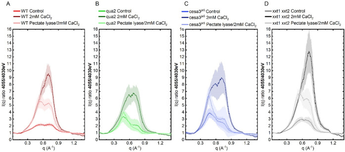
Plant cell walls are abundant sources of materials and energy. Nevertheless, cell wall nanostructure, specifically how pectins interact with cellulose and hemicelluloses to construct a robust and flexible biomaterial, is poorly understood. X-ray scattering measurements are minimally invasive and can reveal ultrastructural, compositional, and physical properties of materials. Resonant X-ray scattering takes advantage of compositional differences by tuning the energy of the incident X-ray to absorption edges of specific elements in a material. Using Tender Resonant X-ray Scattering (TReXS) at the calcium K-edge to study hypocotyls of the model plant, Arabidopsis thaliana, we detected distinctive Ca features that we hypothesize correspond to previously unreported Ca-Homogalacturonan (Ca-HG) nanostructures. When Ca-HG structures were perturbed by chemical and enzymatic treatments, cellulose microfibrils were also rearranged. Moreover, Ca-HG nanostructure was altered in mutants with abnormal cellulose, pectin, or hemicellulose content. Our results indicate direct structural interlinks between components of the plant cell wall at the nanoscale and reveal mechanisms that underpin both the structural integrity of these components and the molecular architecture of the plant cell wall.


Conflict of interest statement

The authors declare no competing financial interest.

Figures

Figure 1
Figure 1
Tender resonant X-ray scattering reveals Ca dependent nanostructure in A. thaliana hypocotyls. (A) Resonant scattering at the Ca K-edge allows distinction of Ca and Ca-cross-linked pectin from other cell wall components. (B) NEXAFS profile of WT hypocotyls at the Ca K-edge with pre-edge energies highlighted in gray and the Ca K-edge in red. (C) I(q) TReXS WAXS profiles at 4030, 4040, 4050, 4055, and 4075 eV. (D) Scattering contrast calculated for cell wall components, vacuum and Ca, normalized to 4030 eV (upper part) that shows Ca structures and 4050 eV (lower part) that deconvolute scattering contrast between Ca within carbon compounds (e.g., Ca-pectins) or Ca-pectin structures in cell wall components background. (E) Composite scattering contrast predictions and total scattering intensity (TSI between 0.2 and 1 A–1) for WT control and Ca-pectin gel samples. (F, G) I(q) 4055/4030 eV ratio and 4075/4050 eV for WT control, CaCl2 and calcium cross-linked pectin dry gel, and hypocotyls treated with CDTA (pectin extraction). Shaded color above and under curves shows standard deviation of n > 9 measurements from 3 independent samples made of >15 dry hypocotyls.
Figure 2
Figure 2
Ca-pectin structure is perturbed in wall component-deficient mutants. (A) I(q) 4055/4030 eV ratios for WT (Ca and pectate lyase treated), (B) I(q) 4055/4030 eV ratios for qua2 pectin deficient mutant (Ca and pectate lyase treated), (C) I(q) 4055/4030 eV ratios for cesa3je5 cellulose-deficient (Ca and pectate lyase treated), and (D) I(q) 4055/4030 eV ratios for the xxt1 xxt2 xyloglucan-deficient mutant (Ca and pectate lyase treated). Shaded color above and under curves shows standard deviation of n > 9 measurements from 3 independent samples made of >15 dry hypocotyls.
Figure 3
Figure 3
Modification of Ca-pectin in native walls affects cellulose microfibril spacing and arrangement. (A) Mean spacing between cellulose microfibrils in WT, qua2, cesa3je5, and xxt1 xxt2 plants under control, Ca, and pectate lyase treatment. (B) Integration along azimuthal angle of SAXS scattering images of WT, qua2, cesa3je5, and xxt1 xxt2 under control, Ca, and pectate lyase treatment. (C) Full width half-maximum (fwhm) of the main peak from SAXS scattering profiles obtained by integrating 2D data along the azimuthal angle for WT, qua2, cesa3je5, and xxt1 xxt2 under control, Ca, and pectate lyase treatment. Different letters (a, b, c) above boxes indicate statistically distinct groups, measured by one-way ANOVA with Tukey HSD, p < 0.05.
Figure 4
Figure 4
Ca-pectin structure with shorter spacing is anisotropic along the longitudinal axis of hypocotyls and microfibrils. (A–C) Scattering images of 4055/4030 eV ratio of WT control, Ca and pectate lyase treated samples, (A’–C’) I(q) 4055/4030 eV ratio profiles integrated at azimuthal angles of 0–25° (red) and 100–75° (blue); data correspond to scattering images A-C.
Figure 5
Figure 5
Graphical interpretation of results. Proposal of the Ca-pectin structure in WT hypocotyl primary cell wall after chemical treatments and its interaction with cellulose structure based on results of Ca-pectin structural changes, cellulose microfibril (CMF) spacing, and CMF bundle alignment in wall component-deficient mutants and following chemical/enzymatic treatments.

References

    1. Wang Y.; Jiao Y. Cellulose Microfibril-Mediated Directional Plant Cell Expansion: Gas and Brake. Mol. Plant 2020, 13 (12), 1670–1672. 10.1016/j.molp.2020.10.010. - DOI - PubMed
    1. Balat M.; Ayar G. Biomass energy in the world, use of biomass and potential trends. Energy sources 2005, 27 (10), 931–940. 10.1080/00908310490449045. - DOI
    1. Rongpipi S.; Ye D.; Gomez E. D.; Gomez E. W. Progress and Opportunities in the Characterization of Cellulose - An Important Regulator of Cell Wall Growth and Mechanics. Front Plant Sci. 2019, 9, 1894.10.3389/fpls.2018.01894. - DOI - PMC - PubMed
    1. Altartouri B.; Bidhendi A. J.; Tani T.; Suzuki J.; Conrad C.; Chebli Y.; Liu N.; Karunakaran C.; Scarcelli G.; Geitmann A. Pectin chemistry and cellulose crystallinity govern pavement cell morphogenesis in a multi-step mechanism. Plant Physiol. 2019, 181 (1), 127–141. 10.1104/pp.19.00303. - DOI - PMC - PubMed
    1. Dehors J.; Mareck A.; Kiefer-Meyer M. C.; Menu-Bouaouiche L.; Lehner A.; Mollet J. C. Evolution of Cell Wall Polymers in Tip-Growing Land Plant Gametophytes: Composition, Distribution, Functional Aspects and Their Remodeling. Front Plant Sci. 2019, 10, 441.10.3389/fpls.2019.00441. - DOI - PMC - PubMed

LinkOut - more resources