. 2023 Dec 28:48:102328.
doi: 10.1016/j.jcot.2023.102328. eCollection 2024 Jan.

Chronic lateral ankle ligament instability - Current evidence and recent management advances

Affiliations

Chronic lateral ankle ligament instability - Current evidence and recent management advances

Choon Chiet Hong et al. J Clin Orthop Trauma. .

Erratum in

Abstract

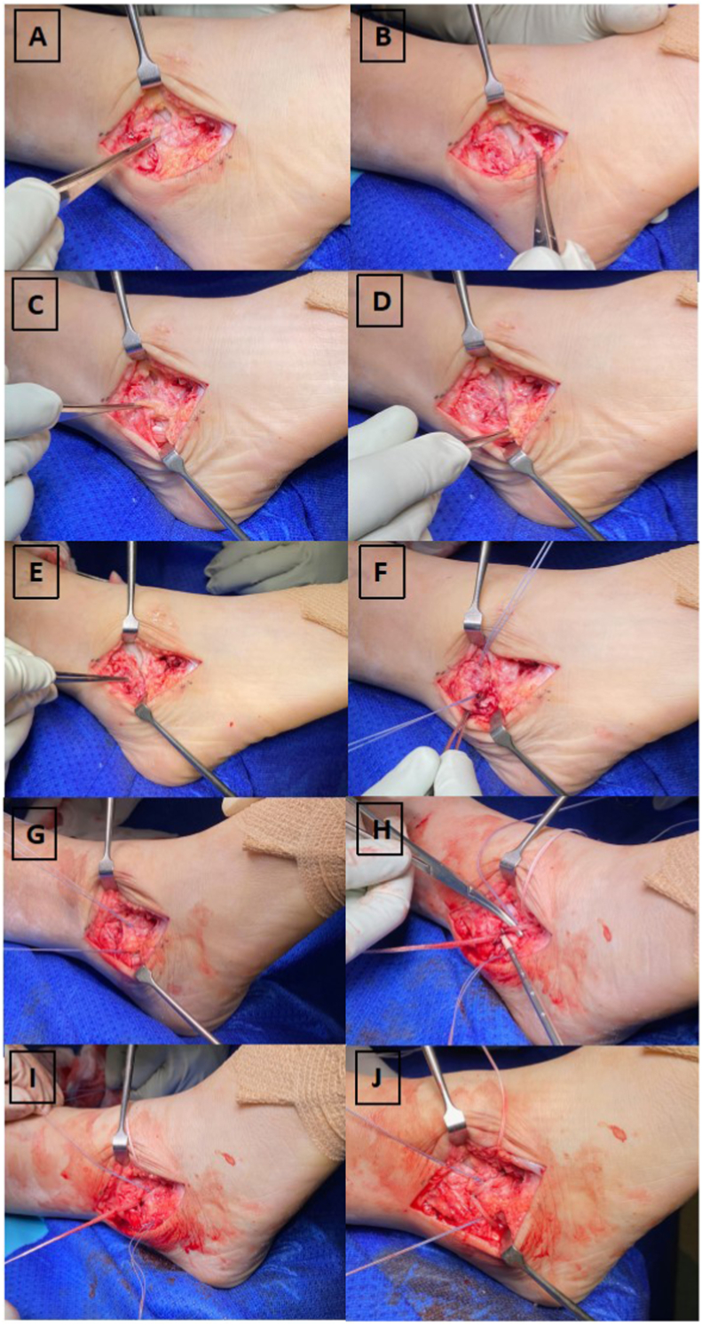
Lateral ankle sprain is a common injury with a substantial negative impact on physical function, quality of life and health economic burden. Chronic lateral ankle instability (CLAI) as a sequela of lateral ankle sprain can lead to the development of posttraumatic ankle osteoarthritis in the long term. In this article, we explore the epidemiology, burden and definition of CLAI for the appropriate clinical assessment and imaging evaluation of patients with lateral ankle sprain and CLAI. Following that, recent advances and evidence on management of CLAI is critically distilled and summarized.


Conflict of interest statement

The authors declare the following financial interests/personal relationships which may be considered as potential competing interests: James Calder reports a relationship with Arthrex that includes: speaking and lecture fees.

Figures

Fig. 1
Fig. 1
1A illustrates the anterior drawer test with the ankle in plantarflexion and the dominant hand holding the distal shin stable while the other hand holds the hindfoot and translating them anteriorly. 1B shows a positive test where there is an anterior translation of the hindfoot.
Fig. 2
Fig. 2
2A and 2B depict the talar tilt test where the ankle is held in a neutral position to lock the talus in the ankle mortise while inverting the hindfoot in relation to the tibia along the coronal plane.
Fig. 3
Fig. 3
3A is the sagittal cut of a normal 2D MRI demonstrating the presence of the superior fascicle of ATFL (white arrow) and the inferior fascicle of ATFL (yellow arrow) connected to the CFL via the arciform fibers (red arrow). 3B is the same sagittal cut but taken 1 slice more medial illustrating the superior (white arrow) and inferior (yellow arrow) fascicles inserting to the lateral border of the talar body. (For interpretation of the references to colour in this figure legend, the reader is referred to the web version of this article.)
Fig. 4
Fig. 4
4A and 4B depict the ATFL ligament and its insertion on the fibula footprint while 4C and 4D demonstrate the CFL ligament and its insertion on the fibula footprint which is inferior to the ATFL while it runs deep to the peroneal tendons inserting to the calcaneus. 4E shows the location of the obscure tubercle which is an important landmark differentiating the insertion of ATFL and CFL. 4F illustrates the insertion of suture anchors at the ATFL and CFL footprint respectively where ATFL is superior to the obscure tubercle while CFL is inferior to it. 4G shows the modified Brostrom-Gould repair where both ATFL and CFL were shortened and repaired to the fibula footprints using suture anchors. 4H demonstrates the use of a synthetic suture tape for augmentation over the ATFL repair highlighting the hemostat under the suture tape to prevent overtightening (check rein) during the final tightening of the suture tape construct. 4I and 4J depicts the extension of the suture tape augmentation for the CFL repair as well. One should take note that the suture tape inserts at the obscure tubercle on the fibula in between both the primary ATFL and CFL repair footprint.
Fig. 5
Fig. 5
5A to 5C illustrates the torn ATFL from the fibula with synovitis in the lateral gutter followed by debridement and preparation of the footprint. 5D shows the ATFL ligament held with grasping sutures. 5E depicts the drilling of the suture anchor drill hole. 5F and 5G demonstrates the introduction of the knotless suture anchor and insertion of the suture anchor into the drill hole while bringing the entire remnant ATFL to the fibula footprint closing the redundant lateral gutter space. 5H shows the probing of the final repair construct and its stability.

References

    1. Herzog M.M., Kerr Z.Y., Marshall S.W., Wikstrom E.A. Epidemiology of ankle sprains and chronic ankle instability. J Athl Train. 2019 Jun;54(6):603–610. - PMC - PubMed
    1. Waterman B.R., Owens B.D., Davey S., Zacchilli M.A., Belmont P.J., Jr. The epidemiology of ankle sprains in the United States. J Bone Joint Surg Am. 2010 Oct 6;92(13):2279–2284. - PubMed
    1. Cooke M.W., Lamb S.E., Marsh J., Dale J. A survey of current consultant practice of treatment of severe ankle sprains in emergency departments in the United Kingdom. Emerg Med J. 2003 Nov;20(6):505–507. - PMC - PubMed
    1. Lamb S.E., Marsh J.L., Hutton J.L., Nakash R., Cooke M.W. Collaborative Ankle Support Trial (CAST Group). mechanical supports for acute, severe ankle sprain: a pragmatic, multicentre, randomised controlled trial. Lancet. 2009 Feb 14;373(9663):575–581. - PubMed
    1. Gribble P.A., Bleakley C.M., Caulfield B.M., et al. Evidence review for the 2016 International Ankle Consortium consensus statement on the prevalence, impact and long-term consequences of lateral ankle sprains. Br J Sports Med. 2016 Dec;50(24):1496–1505. - PubMed

LinkOut - more resources