

Transoesophageal echocardiography-guided balloon-assisted percutaneous closure of a large secundum atrial septal defect in a pregnant woman: a case report
- PMID: 38274706
- PMCID: PMC10810588
- DOI: 10.1093/ehjcr/ytae014
Transoesophageal echocardiography-guided balloon-assisted percutaneous closure of a large secundum atrial septal defect in a pregnant woman: a case report
Abstract
Background: According to the 2018 European Society of Cardiology guidelines, atrial septal defect (ASD) closure can be performed during pregnancy but is rarely indicated. In this case, we demonstrate the viability of percutaneous balloon-assisted ASD closure without fluoroscopy in a pregnant woman.
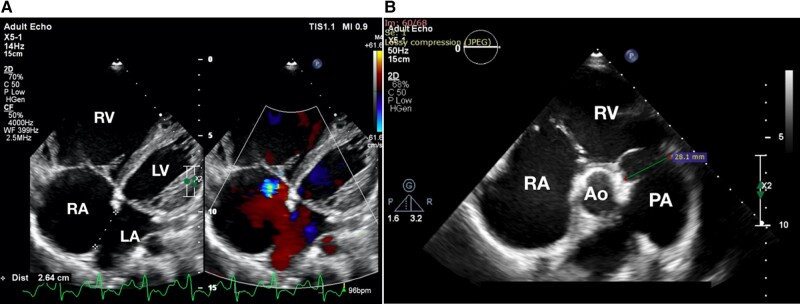
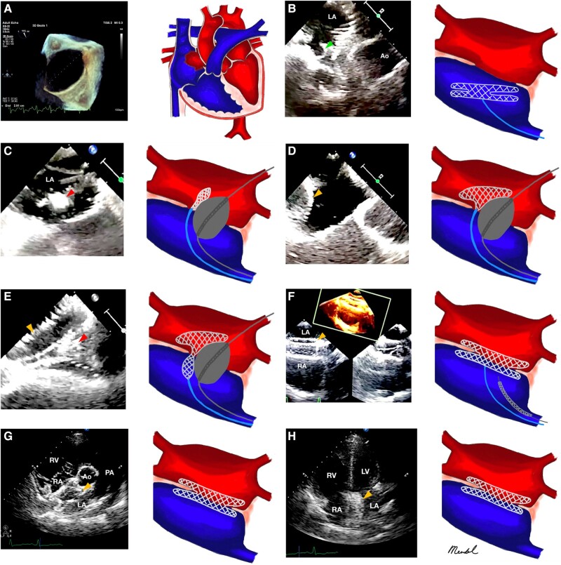
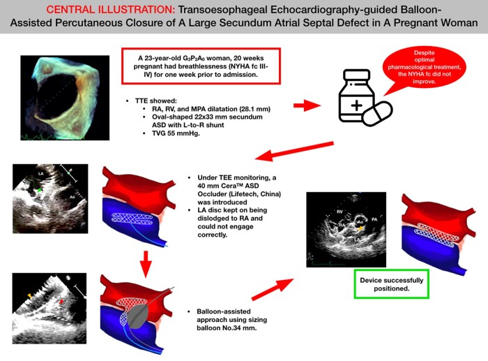
Case summary: A 23-year-old G3P2A0 woman who was 20 weeks pregnant had primary complaints of breathlessness [New York Heart Association functional class (NYHA fc) III and IV] for 1 week prior to admission. A transthoracic echocardiography showed a dilatation of the right atrium (RA), a dilated right ventricle, a dilated main pulmonary artery (28.1 mm), and an oval-shaped 22 × 33 mm-sized secundum ASD with a left-to-right shunt. Despite optimal pharmacological treatment, the NYHA fc persisted. Under transoesophageal echocardiography monitoring, we introduced a 40 mm Cera™ ASD Occluder (Lifetech, China) via the delivery sheath. The device was deployed in the usual position; however, despite numerous placement adjustments, the left atrium disc kept getting dislodged to the RA and could not engage correctly. Therefore, we decided to use a balloon-assisted approach using a sizing balloon of No. 34 mm. The device was successfully positioned, and a wiggle test was conducted to make sure that the device remained stable. The patient was able to give birth to the child normally several months later.
Discussion: Despite the fact that pregnant women with ASD receive a very low dose of radiation, it is nevertheless recommended to avoid radiation because this demographic is particularly vulnerable to it. It is possible to treat a large ASD in pregnant women with a successful balloon-assisted approach.
Keywords: Atrial septal defect; Balloon-assisted; Case report; Non-fluoroscopy; Occluder; Pregnant.
© The Author(s) 2024. Published by Oxford University Press on behalf of the European Society of Cardiology.
Conflict of interest statement
Conflict of interest: None declared.
Figures
Similar articles
-
New modified balloon-assisted technique to provide appropriate deployment in the closure of large secundum atrial septal defect using amplatzer septal occluder in children.J Invasive Cardiol. 2014 Nov;26(11):597-602. J Invasive Cardiol. 2014. PMID: 25364001
-
Transcatheter closure of secundum atrial septal defect using Cocoon septal occluder: immediate and long-term results.Egypt Heart J. 2022 Aug 13;74(1):59. doi: 10.1186/s43044-022-00298-2. Egypt Heart J. 2022. PMID: 35962873 Free PMC article.
-
Clinical results of large secundum atrial septal defect closure in adult using percutaneous transcatheter Cocoon atrial septal occluder.J Med Assoc Thai. 2013 Sep;96(9):1127-34. J Med Assoc Thai. 2013. PMID: 24163987
-
[Percutaneous closure of a very large atrial septal defect: a case report and literature review].G Ital Cardiol (Rome). 2018 Oct;19(10):563-567. doi: 10.1714/2978.29842. G Ital Cardiol (Rome). 2018. PMID: 30281044 Review. Italian.
-
Transcatheter Closure of Atrial Septal Defect: A Review of Currently Used Devices.Cureus. 2023 Jun 8;15(6):e40132. doi: 10.7759/cureus.40132. eCollection 2023 Jun. Cureus. 2023. PMID: 37425612 Free PMC article. Review.
Cited by
-
Safety and Efficacy of Zero Fluoroscopy Patent Ductus Arteriosus Closure in Comparison to the Standardized Fluoroscopy-Guided Procedure: A Systematic Review and Meta-Analysis.Curr Cardiol Rev. 2025;21(5):6-14. doi: 10.2174/011573403X338573241101092849. Curr Cardiol Rev. 2025. PMID: 39901688
-
Echocardiography-guided percutaneous closure of oval-shaped secundum atrial septal defects.BMC Cardiovasc Disord. 2024 Oct 3;24(1):534. doi: 10.1186/s12872-024-04165-7. BMC Cardiovasc Disord. 2024. PMID: 39363250 Free PMC article.
-
Expanding role of absolute zero fluoroscopy atrial septal defect closure: a single-center experience.Front Cardiovasc Med. 2025 Apr 4;12:1430555. doi: 10.3389/fcvm.2025.1430555. eCollection 2025. Front Cardiovasc Med. 2025. PMID: 40255341 Free PMC article.
References
-
- Baumgartner H, Backer JD, Babu-Narayan SV, Budts W, Chessa M, Diller GP, et al. . 2020 ESC guidelines for the management of adult congenital heart disease. Eur Heart J 2021;42:563–645. - PubMed
-
- Stout KK, Daniels CJ, Aboulhosn JA, Bozkurt B, Broberg CS, Colman JM. 2018 AHA/ACC guideline for the management of adults with congenital heart disease: a report of the American College of Cardiology/American Heart Association task force on clinical practice guidelines. Circulation 2019;139:e698–e800. - PubMed
-
- Regitz-Zagrosek V, Roos-Hesselink JW, Bauersachs J, Blomstrom-Lundqvist C, Cifkova R, Bonis MD, et al. . 2018 ESC guidelines for the management of cardiovascular diseases during pregnancy. Eur Heart J 2018;39:3165–3241. - PubMed
Publication types
LinkOut - more resources
Full Text Sources