. 2024 Jan 11:14:1263352.
doi: 10.3389/fimmu.2023.1263352. eCollection 2023.

Impaired macrophage and memory T-cell responses to Bacillus Calmette-Guerin nonpolar lipid extract

Affiliations

Impaired macrophage and memory T-cell responses to Bacillus Calmette-Guerin nonpolar lipid extract

Alice Sarno et al. Front Immunol. .

Abstract

Introduction: The attenuation of BCG has led to the loss of not only immunogenic proteins but also lipid antigens.

Methods: Thus, we compared the macrophage and T-cell responses to nonpolar lipid extracts harvested from BCG and Mycobacterium tuberculosis (Mtb) to better understand the role of BCG lipids in the already known diminished responses of the vaccine strain.

Results: Relative to Mtb, nonpolar lipid extract from BCG presented a reduced capacity to trigger the expression of the genes encoding TNF, IL-1b, IL-6 and IL-10 in RAW 264.7 macrophages. Immunophenotyping of PBMCs isolated from healthy individuals revealed that lipids from both BCG and Mtb were able to induce an increased frequency of CD4+ and CD8+ T cells, but only the lipid extract from Mtb enhanced the frequency of CD4-CD8-double-negative, γσ+, CD4+HLA-DR+, and γσ+HLA-DR+ T cells relative to the nonstimulated control. Interestingly, only the Mtb lipid extract was able to increase the frequency of CD4+ memory (CD45RO+) T cells, whereas the BCG lipid extract induced a diminished frequency of CD4+ central memory (CD45RO+CCR7-) T cells after 48 h of culture compared to Mtb.

Discussion: These findings show that the nonpolar lipids of the BCG bacilli presented diminished ability to trigger both proinflammatory and memory responses and suggest a potential use of Mtb lipids as adjuvants to increase the BCG vaccine efficacy.

Keywords: Bacillus Calmette-Guerin; Mycobacterium tuberculosis; macrophage gene expression; memory T-cell responses; nonpolar lipid extracts.


Conflict of interest statement

The authors declare that the research was conducted in the absence of any commercial or financial relationships that could be construed as a potential conflict of interest.

Figures

Figure 1
Figure 1
RT‒qPCR analyses of (A) IL-1β, (B) IL-6, (C) TNF, and (D) IL-10 after 2 h, 12 h, 24 h, and 72 h of cell exposure to BCG (orange) and Mtb (blue) lipid extracts. Data represent the mean fold-change difference between BCG and Mtb relative to untreated control. Gene expression was normalized to glyceraldehyde 3-phosphate dehydrogenase (GAPDH) and β -actin genes. p values were calculated by t test, with ***p <0.001; ****p <0.0001.
Figure 2
Figure 2
Flow cytometry of conventional and nonconventional T cells after 48 h of in vitro culture of PBMCs from healthy individuals with lipid extracts of BCG and Mtb. (A) and (E) Frequencies of CD4+ and CD4+ HLA-DR+ T cells. (B) and (F) Frequencies of CD8+ and CD8+ HLA-DR+ T cells. (C) and (G) Frequencies of CD4-CD8- DN and CD4-CD8- DN HLA-DR+ T cells. (D) and (H) Frequencies of γδ+ and γδ+ HLA-DR+ T cells. Normal distribution was determined by the Shapiro‒Wilk test. p values for normal distributions were calculated by one-way ANOVA, and p values for nonnormal distributions were calculated by the Kruskal‒Wallis test. *p <0.05; **p <0.01; ***p <0.001. Nonstimulated control (negative control); Phytohemagglutinin, PHA (positive control).
Figure 3
Figure 3
Flow cytometry of memory T cells after 48 h of in vitro culture of PBMCs from healthy individuals with lipid extracts of BCG and Mtb. (A) and (B) Frequencies of CD4+ and CD8+ naïve T cells (CD45RA+). (C) and (D) Frequencies of CD4+ and CD8+ memory T cells (CD45RO+). (E) and (F) Frequencies of CD4+ and CD8+ central memory T cells (CD45RO+ CCR7+). (G) and (H) Frequencies of CD4+ and CD8+ effector memory T cells (CD45RO+ CCR7-). Normal distribution was determined by the Shapiro‒Wilk test. p values for normal distributions were calculated by one-way ANOVA, and p values for nonnormal distributions were calculated by the Kruskal‒Wallis test. **p <0.01; ***p <0.001; ****p <0.0001. Nonstimulated control (negative control); Phytohemagglutinin, PHA (positive control).
Figure 4
Figure 4
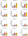
Flow cytometry of CD4+, CD8+ and CD4-CD8- DN T cells producing TNF, IFNγ, IL-2, and IL-17 after 48 h of in vitro culture of PBMCs from healthy individuals with lipid extracts of BCG and Mtb. (A), (B), and (C) MFI of CD4+, CD8+ and CD4-CD8- DN T cells producing TNF. (D), (E), and (F) MFI of CD4+, CD8+ and CD4-CD8- DN T cells producing IFNγ. (G), (H), and (I) MFI of CD4+, CD8+ and CD4-CD8- DN T cells producing IL-2. (J), (K), and (L) MFI of CD4+, CD8+ and CD4-CD8- DN T cells producing IL-17. Normal distribution was determined by the Shapiro‒Wilk test. p values for normal distributions were calculated by one-way ANOVA, and p values for nonnormal distributions were calculated by the Kruskal‒Wallis test. *p <0.05; **p <0.01; ***p <0.001; ****p <0.0001. MFI: Median fluorescence intensity. Nonstimulated control (negative control); Phytohemagglutinin, PHA (positive control).
Figure 5
Figure 5
Concentrations of (A) IFNγ and (B) IL-10 production after 48 h of in vitro culture of PBMCs from healthy individuals with lipid extracts of BCG and Mtb. Normal distribution was determined by the Shapiro‒Wilk test. p values for normal distributions were calculated by one-way ANOVA, and p values for nonnormal distributions were calculated by the Kruskal‒Wallis test. **p <0.01. Nonstimulated control (negative control); Phytohemagglutinin, PHA (positive control).

References

    1. WHO . Global tuberculosis report 2022. Geneva: World Health Organization. (2022).
    1. Abubakar I, Pimpin L, Ariti C, Beynon R, Mangtani P, Sterne J, et al. . Systematic review and meta-analysis of the current evidence on the duration of protection by bacillus Calmette–Guérin vaccination against tuberculosis. Health Technol Assess (2013) 17:. doi: 10.3310/hta17370 - DOI - PMC - PubMed
    1. Mangtani P, Abubakar I, Ariti C, Beynon R, Pimpin L, Fine PEM, et al. . Protection by BCG vaccine against tuberculosis: A systematic review of randomized controlled trials. Clin Infect Dis (2014) 58:470–80. doi: 10.1093/cid/cit790 - DOI - PubMed
    1. Roy A, Eisenhut M, Harris RJ, Rodrigues LC, Sridhar S, Habermann S, et al. . Effect of BCG vaccination against Mycobacterium tuberculosis infection in children: systematic review and meta-analysis. BMJ (2014) 349:1–11. doi: 10.1136/BMJ.G4643 - DOI - PMC - PubMed
    1. Hayashi D, Takii T, Fujiwara N, Fujita Y, Yano I, Yamamoto S, et al. . Comparable studies of immunostimulating activities in vitro among Mycobacterium bovis bacillus Calmette-Guérin (BCG) substrains. FEMS Immunol Med Microbiol (2009) 56:116–28. doi: 10.1111/j.1574-695X.2009.00559.x - DOI - PubMed

Publication types