

The pathogenesis of blepharospasm
- PMID: 38274886
- PMCID: PMC10808626
- DOI: 10.3389/fneur.2023.1336348
The pathogenesis of blepharospasm
Abstract
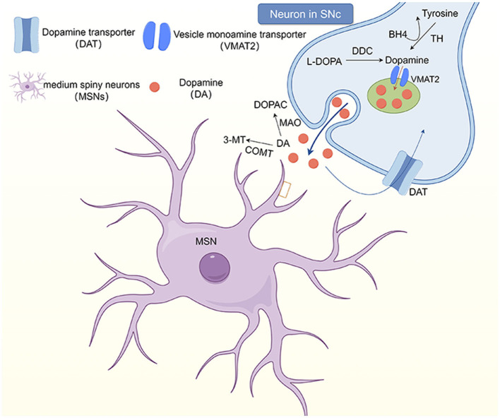
Blepharospasm is a focal dystonia characterized by involuntary tetanic contractions of the orbicularis oculi muscle, which can lead to functional blindness and loss of independent living ability in severe cases. It usually occurs in adults, with a higher incidence rate in women than in men. The etiology and pathogenesis of this disease have not been elucidated to date, but it is traditionally believed to be related to the basal ganglia. Studies have also shown that this is related to the decreased activity of inhibitory neurons in the cerebral cortex caused by environmental factors and genetic predisposition. Increasingly, studies have focused on the imbalance in the regulation of neurotransmitters, including dopamine, serotonin, and acetylcholine, in blepharospasm. The onset of the disease is insidious, and the misdiagnosis rate is high based on history and clinical manifestations. This article reviews the etiology, epidemiological features, and pathogenesis of blepharospasm, to improve understanding of the disease by neurologists and ophthalmologists.
Keywords: basal ganglia; blepharospasm; cerebellum; dopamine; dystonia; neurotransmitters; pathogenesis; serotonin.
Copyright © 2024 Zhu, Meng, Zhang, Xie, Sun and Hou.
Conflict of interest statement
The authors declare that the research was conducted in the absence of any commercial or financial relationships that could be construed as a potential conflict of interest.
Figures
References
-
- Jahngir MU, Ameer MA, Patel BC. Meige syndrome. In: StatPearls. Treasure Island, FL: StatPearls Publishing, L. L. C. Copyright, ©; (2023). - PubMed
Publication types
LinkOut - more resources
Full Text Sources